6QH2 | pdb_00006qh2

Solution NMR ensemble for a chimeric KH-S1 domain construct of exosomal polynucleotide phosphrylase at 298K compiled using the CoMAND method


Experimental Data Snapshot

  • Method: SOLUTION NMR
  • Conformers Calculated: 10000 
  • Conformers Submitted: 20 
  • Selection Criteria: back calculated data agree with experimental NOESY spectrum 

wwPDB Validation 3D Report Full Report

Validation slider image for 6QH2

This is version 1.2 of the entry. See complete history

Literature

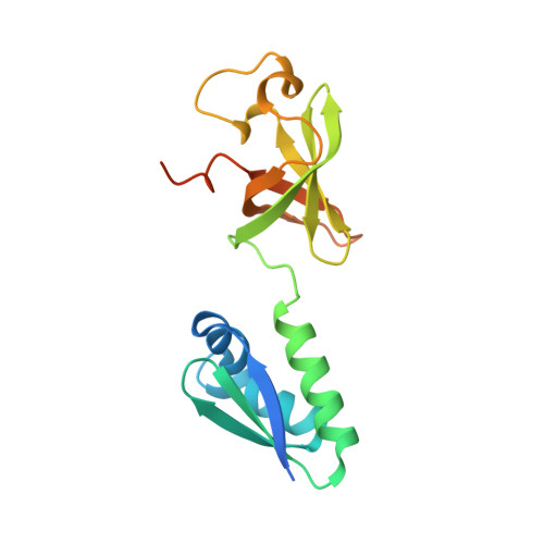
Mapping Local Conformational Landscapes of Proteins in Solution.

ElGamacy, M.Riss, M.Zhu, H.Truffault, V.Coles, M.

(2019) Structure 27: 853-865.e5

  • DOI: https://doi.org/10.1016/j.str.2019.03.005
  • Primary Citation Related Structures: 
    6QF8, 6QFP, 6QH2

  • PubMed Abstract: 

    The ability of proteins to adopt multiple conformational states is essential to their function, and elucidating the details of such diversity under physiological conditions has been a major challenge. Here we present a generalized method for mapping protein population landscapes by NMR spectroscopy. Experimental NOESY spectra are directly compared with a set of expectation spectra back-calculated across an arbitrary conformational space. Signal decomposition of the experimental spectrum then directly yields the relative populations of local conformational microstates. In this way, averaged descriptions of conformation can be eliminated. As the method quantitatively compares experimental and expectation spectra, it inherently delivers an R factor expressing how well structural models explain the input data. We demonstrate that our method extracts sufficient information from a single 3D NOESY experiment to perform initial model building, refinement, and validation, thus offering a complete de novo structure determination protocol.


  • Organizational Affiliation
    • Department of Protein Evolution, Max Planck Institute for Developmental Biology, Max-Planck-Ring 5, 72076 Tübingen, Germany.

Macromolecule Content 

  • Total Structure Weight: 18.4 kDa 
  • Atom Count: 1,094 
  • Modeled Residue Count: 142 
  • Deposited Residue Count: 171 
  • Unique protein chains: 1

Macromolecules

Find similar proteins by:|  3D Structure
Entity ID: 1
MoleculeChains  Sequence LengthOrganismDetailsImage
Polyribonucleotide nucleotidyltransferase171Escherichia coli SE11Mutation(s): 0 
Gene Names: pnpECSE_3450
EC: 2.7.7.8
UniProt
Find proteins for P05055 (Escherichia coli (strain K12))
Explore P05055 
Go to UniProtKB:  P05055
Entity Groups
Sequence Clusters30% Identity50% Identity70% Identity90% Identity95% Identity100% Identity
UniProt GroupP05055
Sequence Annotations
Expand
Reference Sequence

Experimental Data & Validation

Experimental Data

  • Method: SOLUTION NMR
  • Conformers Calculated: 10000 
  • Conformers Submitted: 20 
  • Selection Criteria: back calculated data agree with experimental NOESY spectrum 

Structure Validation

View Full Validation Report


Entry History 

Deposition Data

Revision History  (Full details and data files)

  • Version 1.0: 2019-04-10
    Type: Initial release
  • Version 1.1: 2019-05-15
    Changes: Data collection, Database references
  • Version 1.2: 2024-05-15
    Changes: Data collection, Database references