6QFE | pdb_00006qfe

Crystal Structure of Human Kallikrein 5 in complex with GSK144


Experimental Data Snapshot

  • Method: X-RAY DIFFRACTION
  • Resolution: 1.67 Å
  • R-Value Free: 
    0.242 (Depositor), 0.240 (DCC) 
  • R-Value Work: 
    0.211 (Depositor), 0.212 (DCC) 
  • R-Value Observed: 
    0.213 (Depositor) 

Starting Model: experimental
View more details

wwPDB Validation 3D Report Full Report

Validation slider image for 6QFE

Ligand Structure Quality Assessment 


This is version 2.2 of the entry. See complete history

Literature

Evaluation of a crystallographic surrogate for kallikrein 5 in the discovery of novel inhibitors for Netherton syndrome.

Thorpe, J.H.Edgar, E.V.Smith, K.J.Lewell, X.Q.Rella, M.White, G.V.Polyakova, O.Nassau, P.Walker, A.L.Holmes, D.S.Pearce, A.C.Wang, Y.Liddle, J.Hovnanian, A.

(2019) Acta Crystallogr F Struct Biol Commun 75: 385-391

  • DOI: https://doi.org/10.1107/S2053230X19003169
  • Primary Citation Related Structures: 
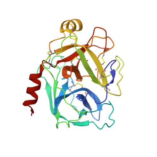
    6QFE, 6QFF, 6QFG, 6QFH

  • PubMed Abstract: 

    The inhibition of kallikrein 5 (KLK5) has been identified as a potential strategy for treatment of the genetic skin disorder Netherton syndrome, in which loss-of-function mutations in the SPINK5 gene lead to down-regulation of the endogenous inhibitor LEKTI-1 and profound skin-barrier defects with severe allergic manifestations. To aid in the development of a medicine for this target, an X-ray crystallographic system was developed to facilitate fragment-guided chemistry and knowledge-based drug-discovery approaches. Here, the development of a surrogate crystallographic system in place of KLK5, which proved to be challenging to crystallize, is described. The biochemical robustness of the crystallographic surrogate and the suitability of the system for the study of small nonpeptidic fragments and lead-like molecules are demonstrated.


  • Organizational Affiliation
    • GlaxoSmithKline, Medicinal Research Centre, Gunnels Wood Road, Stevenage SG1 2NY, England.

Macromolecule Content 

  • Total Structure Weight: 52.52 kDa 
  • Atom Count: 4,083 
  • Modeled Residue Count: 454 
  • Deposited Residue Count: 454 
  • Unique protein chains: 1

Macromolecules

Find similar proteins by:|  3D Structure
Entity ID: 1
MoleculeChains  Sequence LengthOrganismDetailsImage
Kallikrein-5
A, B
227Homo sapiensMutation(s): 0 
Gene Names: KLK5SCTEUNQ570/PRO1132
EC: 3.4.21
UniProt & NIH Common Fund Data Resources
Find proteins for Q9Y337 (Homo sapiens)
Explore Q9Y337 
Go to UniProtKB:  Q9Y337
PHAROS:  Q9Y337
GTEx:  ENSG00000167754 
Entity Groups
Sequence Clusters30% Identity50% Identity70% Identity90% Identity95% Identity100% Identity
UniProt GroupQ9Y337
Glycosylation
Glycosylation Sites: 2Go to GlyGen: Q9Y337-1
Sequence Annotations
Expand
Reference Sequence

Oligosaccharides

Help  
Entity ID: 2
MoleculeChains Length2D Diagram GlycosylationD Interactions
2-acetamido-2-deoxy-beta-D-glucopyranose-(1-4)-2-acetamido-2-deoxy-beta-D-glucopyranose
C, D
2N-Glycosylation
Glycosylation Resources
GlyTouCan: G42666HT
GlyCosmos: G42666HT
GlyGen: G42666HT

Small Molecules

Ligands 3 Unique
IDChains Name / Formula / InChI Key2D Diagram3D Interactions
J08
(Subject of Investigation/LOI)

Query on J08


Download:Ideal Coordinates CCD File
G [auth A],
J [auth B]
4-[(5-phenyl-1~{H}-imidazol-2-yl)methylamino]-2-(pyridin-3-ylmethoxy)benzenecarboximidamide
C23 H22 N6 O
ZDLCAMFQMDNCSU-UHFFFAOYSA-N
NAG

Query on NAG


Download:Ideal Coordinates CCD File
E [auth A],
H [auth B]
2-acetamido-2-deoxy-beta-D-glucopyranose
C8 H15 N O6
OVRNDRQMDRJTHS-FMDGEEDCSA-N
NA

Query on NA


Download:Ideal Coordinates CCD File
F [auth A],
I [auth B]
SODIUM ION
Na
FKNQFGJONOIPTF-UHFFFAOYSA-N

Experimental Data & Validation

Experimental Data

  • Method: X-RAY DIFFRACTION
  • Resolution: 1.67 Å
  • R-Value Free:  0.242 (Depositor), 0.240 (DCC) 
  • R-Value Work:  0.211 (Depositor), 0.212 (DCC) 
  • R-Value Observed: 0.213 (Depositor) 
Space Group: C 1 2 1
Unit Cell:
Length ( Å )Angle ( ˚ )
a = 126.916α = 90
b = 83.84β = 116.23
c = 62.714γ = 90
Software Package:
Software NamePurpose
XDSdata reduction
Aimlessdata scaling
PHASERphasing
BUSTERrefinement
PDB_EXTRACTdata extraction

Structure Validation

View Full Validation Report


Ligand Structure Quality Assessment 


Entry History 

Deposition Data

  • Released Date: 2019-05-08 
  • Deposition Author(s): Thorpe, J.H.

Revision History  (Full details and data files)

  • Version 1.0: 2019-05-08
    Type: Initial release
  • Version 1.1: 2019-05-15
    Changes: Data collection, Database references
  • Version 2.0: 2020-07-29
    Type: Remediation
    Reason: Carbohydrate remediation
    Changes: Atomic model, Data collection, Derived calculations, Structure summary
  • Version 2.1: 2024-01-24
    Changes: Data collection, Database references, Refinement description, Structure summary
  • Version 2.2: 2024-11-13
    Changes: Structure summary