6QEX | pdb_00006qex

Nanodisc reconstituted human ABCB1 in complex with UIC2 fab and taxol


Experimental Data Snapshot

  • Method: ELECTRON MICROSCOPY
  • Resolution: 3.60 Å
  • Aggregation State: PARTICLE 
  • Reconstruction Method: SINGLE PARTICLE 

wwPDB Validation 3D Report Full Report

Validation slider image for 6QEX

This is version 1.4 of the entry. See complete history

Literature

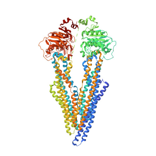
Structural insight into substrate and inhibitor discrimination by human P-glycoprotein.

Alam, A.Kowal, J.Broude, E.Roninson, I.Locher, K.P.

(2019) Science 363: 753-756

  • DOI: https://doi.org/10.1126/science.aav7102
  • Primary Citation Related Structures: 
    6QEE, 6QEX

  • PubMed Abstract: 

    ABCB1, also known as P-glycoprotein, actively extrudes xenobiotic compounds across the plasma membrane of diverse cells, which contributes to cellular drug resistance and interferes with therapeutic drug delivery. We determined the 3.5-angstrom cryo-electron microscopy structure of substrate-bound human ABCB1 reconstituted in lipidic nanodiscs, revealing a single molecule of the chemotherapeutic compound paclitaxel (Taxol) bound in a central, occluded pocket. A second structure of inhibited, human-mouse chimeric ABCB1 revealed two molecules of zosuquidar occupying the same drug-binding pocket. Minor structural differences between substrate- and inhibitor-bound ABCB1 sites are amplified toward the nucleotide-binding domains (NBDs), revealing how the plasticity of the drug-binding site controls the dynamics of the adenosine triphosphate-hydrolyzing NBDs. Ordered cholesterol and phospholipid molecules suggest how the membrane modulates the conformational changes associated with drug binding and transport.


  • Organizational Affiliation
    • Institute of Molecular Biology and Biophysics, ETH Zürich, Otto-Stern-Weg 5, 8093 Zürich, Switzerland.

Macromolecule Content 

  • Total Structure Weight: 199.53 kDa 
  • Atom Count: 13,251 
  • Modeled Residue Count: 1,627 
  • Deposited Residue Count: 1,725 
  • Unique protein chains: 3

Macromolecules

Find similar proteins by:|  3D Structure
Entity ID: 1
MoleculeChains  Sequence LengthOrganismDetailsImage
Multidrug resistance protein 11,280Homo sapiensMutation(s): 1 
Gene Names: ABCB1MDR1PGY1
EC: 7.6.2.2 (PDB Primary Data), 7.6.2.1 (UniProt)
Membrane Entity: Yes 
UniProt & NIH Common Fund Data Resources
Find proteins for P08183 (Homo sapiens)
Explore P08183 
Go to UniProtKB:  P08183
PHAROS:  P08183
GTEx:  ENSG00000085563 
Entity Groups
Sequence Clusters30% Identity50% Identity70% Identity90% Identity95% Identity100% Identity
UniProt GroupP08183
Glycosylation
Glycosylation Sites: 3Go to GlyGen: P08183-1
Sequence Annotations
Expand
Reference Sequence
Find similar proteins by:|  3D Structure
Entity ID: 2
MoleculeChains  Sequence LengthOrganismDetailsImage
UIC2 Fab lightchain220Mus musculusMutation(s): 0 
Find similar proteins by:|  3D Structure
Entity ID: 3
MoleculeChains  Sequence LengthOrganismDetailsImage
UIC2 Fab heavy chain225Mus musculusMutation(s): 0 

Small Molecules

Ligands 4 Unique
IDChains Name / Formula / InChI Key2D Diagram3D Interactions
TA1

Query on TA1


Download:Ideal Coordinates CCD File
W [auth A]TAXOL
C47 H51 N O14
RCINICONZNJXQF-MZXODVADSA-N
3PE

Query on 3PE


Download:Ideal Coordinates CCD File
X [auth A],
Y [auth A]
1,2-Distearoyl-sn-glycerophosphoethanolamine
C41 H82 N O8 P
LVNGJLRDBYCPGB-LDLOPFEMSA-N
CLR

Query on CLR


Download:Ideal Coordinates CCD File
G [auth A]
H [auth A]
I [auth A]
J [auth A]
K [auth A]
G [auth A],
H [auth A],
I [auth A],
J [auth A],
K [auth A],
L [auth A],
M [auth A],
N [auth A],
O [auth A],
P [auth A],
Q [auth A],
R [auth A],
S [auth A],
T [auth A],
U [auth A],
V [auth A]
CHOLESTEROL
C27 H46 O
HVYWMOMLDIMFJA-DPAQBDIFSA-N
NAG

Query on NAG


Download:Ideal Coordinates CCD File
D [auth A],
E [auth A],
F [auth A]
2-acetamido-2-deoxy-beta-D-glucopyranose
C8 H15 N O6
OVRNDRQMDRJTHS-FMDGEEDCSA-N

Experimental Data & Validation

Experimental Data

  • Method: ELECTRON MICROSCOPY
  • Resolution: 3.60 Å
  • Aggregation State: PARTICLE 
  • Reconstruction Method: SINGLE PARTICLE 

Structure Validation

View Full Validation Report


Entry History 

& Funding Information

Deposition Data


Funding OrganizationLocationGrant Number
Swiss National Science FoundationSwitzerland--
European Molecular Biology OrganizationSwitzerland--

Revision History  (Full details and data files)

  • Version 1.0: 2019-02-27
    Type: Initial release
  • Version 1.1: 2019-12-18
    Changes: Other
  • Version 1.2: 2020-07-29
    Type: Remediation
    Reason: Carbohydrate remediation
    Changes: Data collection, Derived calculations, Structure summary
  • Version 1.3: 2021-06-02
    Changes: Structure summary
  • Version 1.4: 2024-11-13
    Changes: Data collection, Database references, Derived calculations, Structure summary