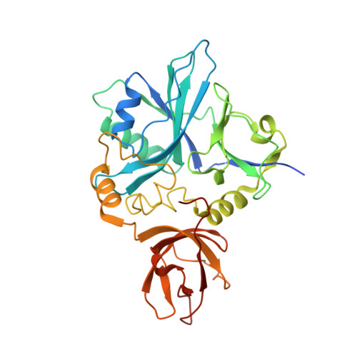
6QE4

Re-refinement of 5OLI human IBA57-I3C


Experimental Data Snapshot

  • Method: X-RAY DIFFRACTION
  • Resolution: 2.30 Å
  • R-Value Free: 0.228 
  • R-Value Work: 0.206 
  • R-Value Observed: 0.208 

wwPDB Validation   3D Report Full Report


Ligand Structure Quality Assessment 


This is version 1.2 of the entry. See complete history

Re-refinement Note

This entry reflects an alternative modeling of the original data in: 6ESR


Literature

In-house high-energy-remote SAD phasing using the magic triangle: how to tackle the P1 low symmetry using multiple orientations of the same crystal of human IBA57 to increase the multiplicity.

Gourdoupis, S.Nasta, V.Ciofi-Baffoni, S.Banci, L.Calderone, V.

(2019) Acta Crystallogr D Struct Biol 75: 317-324

  • DOI: https://doi.org/10.1107/S2059798319000214
  • Primary Citation of Related Structures:  
    6QE3, 6QE4

  • PubMed Abstract: 

    This article describes the approach used to solve the structure of human IBA57 in-house by 5-amino-2,4,6-triiodoisophthalic acid (I3C) high-energy-remote single-wavelength anomalous dispersion (SAD) phasing. Multiple orientations of the same triclinic crystal were exploited to acquire sufficient real data multiplicity for phasing. How the collection of an in-house native data set and its joint use with the I3C derivative through a SIRAS approach decreases the data multiplicity needed by almost 50% is described. Furthermore, it is illustrated that there is a clear data-multiplicity threshold value for success and failure in phasing, and how adding further data does not significantly affect substructure solution and model building. To our knowledge, this is the only structure present in the PDB that has been solved in-house by remote SAD phasing in space group P1 using only one crystal. All of the raw data used, derived from the different orientations, have been uploaded to Zenodo in order to enable software developers to improve methods for data processing and structure solution, and for educational purposes.


  • Organizational Affiliation

    CERM - Magnetic Resonance Center, University of Florence, Via Luigi Sacconi 6, 50019 Sesto Fiorentino (FI), Italy.


Macromolecules
Find similar proteins by:  (by identity cutoff)  |  3D Structure
Entity ID: 1
MoleculeChains Sequence LengthOrganismDetailsImage
Putative transferase CAF17, mitochondrial327Homo sapiensMutation(s): 0 
Gene Names: IBA57C1orf69
EC: 2.1
UniProt & NIH Common Fund Data Resources
Find proteins for Q5T440 (Homo sapiens)
Explore Q5T440 
Go to UniProtKB:  Q5T440
PHAROS:  Q5T440
GTEx:  ENSG00000181873 
Entity Groups  
Sequence Clusters30% Identity50% Identity70% Identity90% Identity95% Identity100% Identity
UniProt GroupQ5T440
Sequence Annotations
Expand
  • Reference Sequence
Experimental Data & Validation

Experimental Data

  • Method: X-RAY DIFFRACTION
  • Resolution: 2.30 Å
  • R-Value Free: 0.228 
  • R-Value Work: 0.206 
  • R-Value Observed: 0.208 
  • Space Group: P 1
Unit Cell:
Length ( Å )Angle ( ˚ )
a = 38.111α = 77.92
b = 43.048β = 75.63
c = 59.281γ = 71.67
Software Package:
Software NamePurpose
XDSdata reduction
XSCALEdata scaling
SHELXDphasing
BUSTERrefinement

Structure Validation

View Full Validation Report


Ligand Structure Quality Assessment 


Entry History 

Deposition Data

Revision History  (Full details and data files)

  • Version 1.0: 2019-03-13
    Type: Initial release
  • Version 1.1: 2019-04-17
    Changes: Data collection, Database references
  • Version 1.2: 2024-05-15
    Changes: Data collection, Database references, Derived calculations