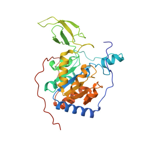
6QCJ

Human Sirt6 in complex with ADP-ribose and the inhibitor catechin gallate


Experimental Data Snapshot

  • Method: X-RAY DIFFRACTION
  • Resolution: 2.01 Å
  • R-Value Free: 0.199 
  • R-Value Work: 0.163 
  • R-Value Observed: 0.165 

Starting Model: experimental
View more details

wwPDB Validation   3D Report Full Report


Ligand Structure Quality Assessment 


This is version 1.1 of the entry. See complete history


Literature

Structural basis for the activation and inhibition of Sirtuin 6 by quercetin and its derivatives.

You, W.Zheng, W.Weiss, S.Chua, K.F.Steegborn, C.

(2019) Sci Rep 9: 19176-19176

  • DOI: https://doi.org/10.1038/s41598-019-55654-1
  • Primary Citation of Related Structures:  
    6QCD, 6QCE, 6QCH, 6QCJ, 6QCN

  • PubMed Abstract: 

    Mammalian Sirtuin 6 (Sirt6) is an NAD + -dependent protein deacylase regulating metabolism and chromatin homeostasis. Sirt6 activation protects against metabolic and aging-related diseases, and Sirt6 inhibition is considered a cancer therapy. Available Sirt6 modulators show insufficient potency and specificity, and even partially contradictory Sirt6 effects were reported for the plant flavone quercetin. To understand Sirt6 modulation by quercetin-based compounds, we analysed their binding and activity effects on Sirt6 and other Sirtuin isoforms and solved crystal structures of compound complexes with Sirt6 and Sirt2. We find that quercetin activates Sirt6 via the isoform-specific binding site for pyrrolo[1,2-a]quinoxalines. Its inhibitory effect on other isoforms is based on an alternative binding site at the active site entrance. Based on these insights, we identified isoquercetin as a ligand that can discriminate both sites and thus activates Sirt6 with increased specificity. Furthermore, we find that quercetin derivatives that inhibit rather than activate Sirt6 exploit the same general Sirt6 binding site as the activators, identifying it as a versatile allosteric site for Sirt6 modulation. Our results thus provide a structural basis for Sirtuin effects of quercetin-related compounds and helpful insights for Sirt6-targeted drug development.


  • Organizational Affiliation

    Department of Biochemistry, University of Bayreuth, 95445, Bayreuth, Germany.


Macromolecules
Find similar proteins by:  (by identity cutoff)  |  3D Structure
Entity ID: 1
MoleculeChains Sequence LengthOrganismDetailsImage
NAD-dependent protein deacetylase sirtuin-6
A, B
302Homo sapiensMutation(s): 0 
Gene Names: SIRT6SIR2L6
EC: 3.5.1 (PDB Primary Data), 2.4.2 (UniProt), 2.3.1 (UniProt), 2.3.1.286 (UniProt)
UniProt & NIH Common Fund Data Resources
Find proteins for Q8N6T7 (Homo sapiens)
Explore Q8N6T7 
Go to UniProtKB:  Q8N6T7
PHAROS:  Q8N6T7
GTEx:  ENSG00000077463 
Entity Groups  
Sequence Clusters30% Identity50% Identity70% Identity90% Identity95% Identity100% Identity
UniProt GroupQ8N6T7
Sequence Annotations
Expand
  • Reference Sequence
Small Molecules
Ligands 5 Unique
IDChains Name / Formula / InChI Key2D Diagram3D Interactions
AR6
Query on AR6

Download Ideal Coordinates CCD File 
C [auth A],
J [auth B]
[(2R,3S,4R,5R)-5-(6-AMINOPURIN-9-YL)-3,4-DIHYDROXY-OXOLAN-2-YL]METHYL [HYDROXY-[[(2R,3S,4R,5S)-3,4,5-TRIHYDROXYOXOLAN-2-YL]METHOXY]PHOSPHORYL] HYDROGEN PHOSPHATE
C15 H23 N5 O14 P2
SRNWOUGRCWSEMX-ZQSHOCFMSA-N
XEG
Query on XEG

Download Ideal Coordinates CCD File 
I [auth A],
P [auth B]
(2R,3S)-2-(3,4-dihydroxyphenyl)-5,7-dihydroxy-3,4-dihydro-2H-chromen-3-yl 3,4,5-trihydroxybenzoate
C22 H18 O10
LSHVYAFMTMFKBA-PZJWPPBQSA-N
SO4
Query on SO4

Download Ideal Coordinates CCD File 
F [auth A]
G [auth A]
H [auth A]
L [auth B]
M [auth B]
F [auth A],
G [auth A],
H [auth A],
L [auth B],
M [auth B],
N [auth B],
O [auth B]
SULFATE ION
O4 S
QAOWNCQODCNURD-UHFFFAOYSA-L
ZN
Query on ZN

Download Ideal Coordinates CCD File 
D [auth A],
K [auth B]
ZINC ION
Zn
PTFCDOFLOPIGGS-UHFFFAOYSA-N
EDO
Query on EDO

Download Ideal Coordinates CCD File 
E [auth A]1,2-ETHANEDIOL
C2 H6 O2
LYCAIKOWRPUZTN-UHFFFAOYSA-N
Experimental Data & Validation

Experimental Data

  • Method: X-RAY DIFFRACTION
  • Resolution: 2.01 Å
  • R-Value Free: 0.199 
  • R-Value Work: 0.163 
  • R-Value Observed: 0.165 
  • Space Group: P 63
Unit Cell:
Length ( Å )Angle ( ˚ )
a = 91.917α = 90
b = 91.917β = 90
c = 144.368γ = 120
Software Package:
Software NamePurpose
REFMACrefinement
XDSdata reduction
XDSdata scaling
PHASERphasing

Structure Validation

View Full Validation Report


Ligand Structure Quality Assessment 


Entry History & Funding Information

Deposition Data


Funding OrganizationLocationGrant Number
Germany--

Revision History  (Full details and data files)

  • Version 1.0: 2019-12-25
    Type: Initial release
  • Version 1.1: 2024-01-24
    Changes: Data collection, Database references, Refinement description