6Q9V | pdb_00006q9v

MSRB3


Experimental Data Snapshot

  • Method: X-RAY DIFFRACTION
  • Resolution: 1.85 Å
  • R-Value Free: 
    0.226 (Depositor), 0.227 (DCC) 
  • R-Value Work: 
    0.182 (Depositor), 0.183 (DCC) 
  • R-Value Observed: 
    0.184 (Depositor) 

Starting Model: experimental
View more details

wwPDB Validation 3D Report Full Report

Validation slider image for 6Q9V

This is version 1.3 of the entry. See complete history

Literature

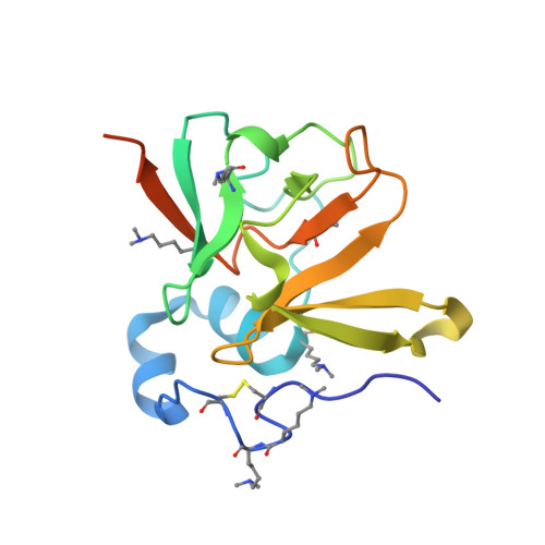
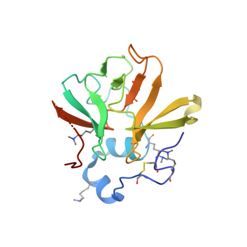
Structure and Electron-Transfer Pathway of the Human Methionine Sulfoxide Reductase MsrB3.

Javitt, G.Cao, Z.Resnick, E.Gabizon, R.Bulleid, N.J.Fass, D.

(2020) Antioxid Redox Signal 33: 665-678

  • DOI: https://doi.org/10.1089/ars.2020.8037
  • Primary Citation Related Structures: 
    6Q9V, 6QA0

  • PubMed Abstract: 

    Aims: The post-translational oxidation of methionine to methionine sulfoxide (MetSO) is a reversible process, enabling the repair of oxidative damage to proteins and the use of sulfoxidation as a regulatory switch. MetSO reductases catalyze the stereospecific reduction of MetSO. One of the mammalian MetSO reductases, MsrB3, has a signal sequence for entry into the endoplasmic reticulum (ER). In the ER, MsrB3 is expected to encounter a distinct redox environment compared with its paralogs in the cytosol, nucleus, and mitochondria. We sought to determine the location and arrangement of MsrB3 redox-active cysteines, which may couple MsrB3 activity to other redox events in the ER. Results: We determined the human MsrB3 structure by using X-ray crystallography. The structure revealed that a disulfide bond near the protein amino terminus is distant in space from the active site. Nevertheless, biochemical assays showed that these amino-terminal cysteines are oxidized by the MsrB3 active site after its reaction with MetSO. Innovation: This study reveals a mechanism to shuttle oxidizing equivalents from the primary MsrB3 active site toward the enzyme surface, where they would be available for further dithiol-disulfide exchange reactions. Conclusion: Conformational changes must occur during the MsrB3 catalytic cycle to transfer oxidizing equivalents from the active site to the amino-terminal redox-active disulfide. The accessibility of this exposed disulfide may help couple MsrB3 activity to other dithiol-disulfide redox events in the secretory pathway.


  • Organizational Affiliation
    • Department of Structural Biology and Weizmann Institute of Science, Rehovot, Israel.

Macromolecule Content 

  • Total Structure Weight: 35.54 kDa 
  • Atom Count: 2,600 
  • Modeled Residue Count: 287 
  • Deposited Residue Count: 317 
  • Unique protein chains: 2

Macromolecules

Find similar proteins by:|  3D Structure
Entity ID: 1
MoleculeChains  Sequence LengthOrganismDetailsImage
Methionine-R-sulfoxide reductase B3160Homo sapiensMutation(s): 0 
Gene Names: MSRB3UNQ1965/PRO4487
EC: 1.8.4.12 (PDB Primary Data), 1.8.4.14 (PDB Primary Data)
UniProt & NIH Common Fund Data Resources
Find proteins for Q8IXL7 (Homo sapiens)
Explore Q8IXL7 
Go to UniProtKB:  Q8IXL7
PHAROS:  Q8IXL7
GTEx:  ENSG00000174099 
Entity Groups
Sequence Clusters30% Identity50% Identity70% Identity90% Identity95% Identity100% Identity
UniProt GroupQ8IXL7
Sequence Annotations
Expand
Reference Sequence
Find similar proteins by:|  3D Structure
Entity ID: 2
MoleculeChains  Sequence LengthOrganismDetailsImage
Methionine-R-sulfoxide reductase B3157Homo sapiensMutation(s): 0 
Gene Names: MSRB3UNQ1965/PRO4487
EC: 1.8.4.12 (PDB Primary Data), 1.8.4.14 (PDB Primary Data)
UniProt & NIH Common Fund Data Resources
Find proteins for Q8IXL7 (Homo sapiens)
Explore Q8IXL7 
Go to UniProtKB:  Q8IXL7
PHAROS:  Q8IXL7
GTEx:  ENSG00000174099 
Entity Groups
Sequence Clusters30% Identity50% Identity70% Identity90% Identity95% Identity100% Identity
UniProt GroupQ8IXL7
Sequence Annotations
Expand
Reference Sequence

Small Molecules

Ligands 3 Unique
IDChains Name / Formula / InChI Key2D Diagram3D Interactions
MPD

Query on MPD


Download:Ideal Coordinates CCD File
E [auth A],
F [auth A],
J [auth B]
(4S)-2-METHYL-2,4-PENTANEDIOL
C6 H14 O2
SVTBMSDMJJWYQN-YFKPBYRVSA-N
SO4

Query on SO4


Download:Ideal Coordinates CCD File
D [auth A],
H [auth B],
I [auth B]
SULFATE ION
O4 S
QAOWNCQODCNURD-UHFFFAOYSA-L
ZN

Query on ZN


Download:Ideal Coordinates CCD File
C [auth A],
G [auth B]
ZINC ION
Zn
PTFCDOFLOPIGGS-UHFFFAOYSA-N
Modified Residues  2 Unique
IDChains TypeFormula2D DiagramParent
CSO
Query on CSO
A
L-PEPTIDE LINKINGC3 H7 N O3 SCYS
MLY
Query on MLY
A
L-PEPTIDE LINKINGC8 H18 N2 O2LYS

Experimental Data & Validation

Experimental Data

  • Method: X-RAY DIFFRACTION
  • Resolution: 1.85 Å
  • R-Value Free:  0.226 (Depositor), 0.227 (DCC) 
  • R-Value Work:  0.182 (Depositor), 0.183 (DCC) 
  • R-Value Observed: 0.184 (Depositor) 
Space Group: P 21 21 21
Unit Cell:
Length ( Å )Angle ( ˚ )
a = 44.025α = 90
b = 65.183β = 90
c = 112.777γ = 90
Software Package:
Software NamePurpose
PHENIXrefinement
xia2data scaling
PHASERphasing
PDB_EXTRACTdata extraction
xia2data reduction

Structure Validation

View Full Validation Report


Entry History 

Deposition Data

Revision History  (Full details and data files)

  • Version 1.0: 2020-01-15
    Type: Initial release
  • Version 1.1: 2020-07-22
    Changes: Database references
  • Version 1.2: 2020-09-09
    Changes: Database references, Derived calculations
  • Version 1.3: 2024-01-24
    Changes: Data collection, Database references, Refinement description