6Q4R | pdb_00006q4r

High-resolution crystal structure of ERAP1 with bound phosphinic transition-state analogue inhibitor


Experimental Data Snapshot

  • Method: X-RAY DIFFRACTION
  • Resolution: 1.60 Å
  • R-Value Free: 
    0.213 (Depositor), 0.215 (DCC) 
  • R-Value Work: 
    0.180 (Depositor), 0.184 (DCC) 
  • R-Value Observed: 
    0.182 (Depositor) 

Starting Model: experimental
View more details

wwPDB Validation 3D Report Full Report

Validation slider image for 6Q4R

Ligand Structure Quality Assessment 


This is version 2.2 of the entry. See complete history

Literature

High-Resolution Crystal Structure of Endoplasmic Reticulum Aminopeptidase 1 with Bound Phosphinic Transition-State Analogue Inhibitor.

Giastas, P.Neu, M.Rowland, P.Stratikos, E.

(2019) ACS Med Chem Lett 10: 708-713

  • DOI: https://doi.org/10.1021/acsmedchemlett.9b00002
  • Primary Citation Related Structures: 
    6Q4R

  • PubMed Abstract: 

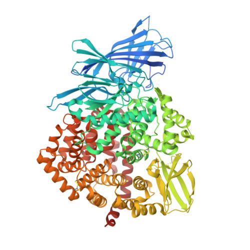
    Endoplasmic reticulum aminopeptidase 1 (ERAP1) is an intracellular enzyme that helps generate peptides presented by Major Histocompatibility Complex Class I (MHC class I) molecules and is an emerging target for immunotherapy applications. Despite almost two decades of research on ERAP1, lack of high-resolution crystal structures has hampered drug-development efforts. By optimizing the protein construct, we obtained a high-resolution (1.60 Å) crystal structure of the closed-conformation of ERAP1 with a potent phosphinic pseudopeptide inhibitor bound in its active site. The structure provides key insight on the mechanism of inhibition as well as selectivity toward homologous enzymes and allows detailed mapping of the internal cavity of the enzyme that accommodates peptide-substrates. Bis-tris propane and malic acid molecules, found bound in pockets in the internal cavity, reveal potential druggable secondary binding sites. The ability to obtain high-resolution crystal structures of ERAP1 removes a major bottleneck in the development of compounds that regulate its activity and will greatly accelerate drug-discovery efforts.


  • Organizational Affiliation
    • National Center for Scientific Research Demokritos, Agia Paraskevi, Athens 15310, Greece.

Macromolecule Content 

  • Total Structure Weight: 108.76 kDa 
  • Atom Count: 8,229 
  • Modeled Residue Count: 855 
  • Deposited Residue Count: 912 
  • Unique protein chains: 1

Macromolecules

Find similar proteins by:|  3D Structure
Entity ID: 1
MoleculeChains  Sequence LengthOrganismDetailsImage
Endoplasmic reticulum aminopeptidase 1,Endoplasmic reticulum aminopeptidase 1912Homo sapiensMutation(s): 0 
Gene Names: ERAP1APPILSARTS1KIAA0525UNQ584/PRO1154
EC: 3.4.11
UniProt & NIH Common Fund Data Resources
Find proteins for Q9NZ08 (Homo sapiens)
Explore Q9NZ08 
Go to UniProtKB:  Q9NZ08
PHAROS:  Q9NZ08
GTEx:  ENSG00000164307 
Entity Groups
Sequence Clusters30% Identity50% Identity70% Identity90% Identity95% Identity100% Identity
UniProt GroupQ9NZ08
Glycosylation
Glycosylation Sites: 4Go to GlyGen: Q9NZ08-1
Sequence Annotations
Expand
Reference Sequence

Oligosaccharides

Help  
Entity ID: 2
MoleculeChains Length2D Diagram GlycosylationD Interactions
2-acetamido-2-deoxy-beta-D-glucopyranose-(1-4)-2-acetamido-2-deoxy-beta-D-glucopyranose
B
2N-Glycosylation
Glycosylation Resources
GlyTouCan: G42666HT
GlyCosmos: G42666HT
GlyGen: G42666HT
Entity ID: 3
MoleculeChains Length2D Diagram GlycosylationD Interactions
beta-D-mannopyranose-(1-4)-2-acetamido-2-deoxy-beta-D-glucopyranose-(1-4)-2-acetamido-2-deoxy-beta-D-glucopyranose
C
3N-Glycosylation
Glycosylation Resources
GlyTouCan: G15407YE
GlyCosmos: G15407YE
GlyGen: G15407YE

Small Molecules

Ligands 9 Unique
IDChains Name / Formula / InChI Key2D Diagram3D Interactions
HJ5
(Subject of Investigation/LOI)

Query on HJ5


Download:Ideal Coordinates CCD File
G [auth A][(1~{R})-1-[[(2~{S})-2-[[(2~{S})-1-azaniumyl-1-oxidanylidene-3-phenyl-propan-2-yl]carbamoyl]pent-4-ynyl]-oxidanyl-phosphoryl]-3-phenyl-propyl]azanium
C24 H32 N3 O4 P
QCPJTEUURKCRNT-BHIFYINESA-P
B3P

Query on B3P


Download:Ideal Coordinates CCD File
H [auth A]2-[3-(2-HYDROXY-1,1-DIHYDROXYMETHYL-ETHYLAMINO)-PROPYLAMINO]-2-HYDROXYMETHYL-PROPANE-1,3-DIOL
C11 H26 N2 O6
HHKZCCWKTZRCCL-UHFFFAOYSA-N
P6G

Query on P6G


Download:Ideal Coordinates CCD File
NA [auth A]HEXAETHYLENE GLYCOL
C12 H26 O7
IIRDTKBZINWQAW-UHFFFAOYSA-N
NAG

Query on NAG


Download:Ideal Coordinates CCD File
D [auth A],
E [auth A]
2-acetamido-2-deoxy-beta-D-glucopyranose
C8 H15 N O6
OVRNDRQMDRJTHS-FMDGEEDCSA-N
PG4

Query on PG4


Download:Ideal Coordinates CCD File
OA [auth A]TETRAETHYLENE GLYCOL
C8 H18 O5
UWHCKJMYHZGTIT-UHFFFAOYSA-N
MLT

Query on MLT


Download:Ideal Coordinates CCD File
MA [auth A]D-MALATE
C4 H6 O5
BJEPYKJPYRNKOW-UWTATZPHSA-N
ZN

Query on ZN


Download:Ideal Coordinates CCD File
F [auth A]ZINC ION
Zn
PTFCDOFLOPIGGS-UHFFFAOYSA-N
EDO

Query on EDO


Download:Ideal Coordinates CCD File
AA [auth A]
BA [auth A]
CA [auth A]
DA [auth A]
EA [auth A]
AA [auth A],
BA [auth A],
CA [auth A],
DA [auth A],
EA [auth A],
FA [auth A],
GA [auth A],
HA [auth A],
I [auth A],
IA [auth A],
J [auth A],
JA [auth A],
K [auth A],
KA [auth A],
L [auth A],
LA [auth A],
M [auth A],
N [auth A],
O [auth A],
P [auth A],
Q [auth A],
R [auth A],
S [auth A],
T [auth A],
U [auth A],
V [auth A],
W [auth A],
X [auth A],
Y [auth A],
Z [auth A]
1,2-ETHANEDIOL
C2 H6 O2
LYCAIKOWRPUZTN-UHFFFAOYSA-N
NA

Query on NA


Download:Ideal Coordinates CCD File
PA [auth A],
QA [auth A],
RA [auth A]
SODIUM ION
Na
FKNQFGJONOIPTF-UHFFFAOYSA-N

Experimental Data & Validation

Experimental Data

  • Method: X-RAY DIFFRACTION
  • Resolution: 1.60 Å
  • R-Value Free:  0.213 (Depositor), 0.215 (DCC) 
  • R-Value Work:  0.180 (Depositor), 0.184 (DCC) 
  • R-Value Observed: 0.182 (Depositor) 
Space Group: P 2 21 21
Unit Cell:
Length ( Å )Angle ( ˚ )
a = 57.683α = 90
b = 116.669β = 90
c = 147.267γ = 90
Software Package:
Software NamePurpose
PHENIXrefinement
XDSdata reduction
XDSdata scaling
PHASERphasing

Structure Validation

View Full Validation Report


Ligand Structure Quality Assessment 


Entry History 

Deposition Data

Revision History  (Full details and data files)

  • Version 1.0: 2019-04-10
    Type: Initial release
  • Version 1.1: 2019-05-29
    Changes: Data collection, Database references
  • Version 2.0: 2020-07-29
    Type: Remediation
    Reason: Carbohydrate remediation
    Changes: Atomic model, Data collection, Derived calculations, Structure summary
  • Version 2.1: 2024-01-24
    Changes: Advisory, Data collection, Database references, Refinement description, Structure summary
  • Version 2.2: 2024-10-16
    Changes: Structure summary