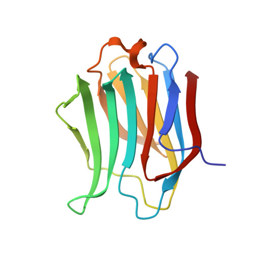
Linear triazole-linked pseudo oligogalactosides as scaffolds for galectin inhibitor development.
Dussouy, C., Kishor, C., Lambert, A., Lamoureux, C., Blanchard, H., Grandjean, C.(2020) Chem Biol Drug Des 96: 1123-1133
- PubMed: 32220037
- DOI: https://doi.org/10.1111/cbdd.13683
- Primary Citation of Related Structures:
6Q0Q, 6Q17 - PubMed Abstract:
Galectins play key roles in numerous biological processes. Their mode of action depends on their localization which can be extracellular, cytoplasmic, or nuclear and is partly mediated through interactions with β-galactose containing glycans. Galectins have emerged as novel therapeutic targets notably for the treatment of inflammatory disorders and cancers. This has stimulated the design of carbohydrate-based inhibitors targeting the carbohydrate recognition domains (CRDs) of the galectins. Pursuing this approach, we reasoned that linear oligogalactosides obtained by straightforward iterative click chemistry could mimic poly-lactosamine motifs expressed at eukaryote cell surfaces which the extracellular form of galectin-3, a prominent member of the galectin family, specifically recognizes. Affinities toward galectin-3 consistently increased with the length of the representative oligogalactosides but without reaching that of oligo-lactosamines. Elucidation of the X-ray crystal structures of the galectin-3 CRD in complex with a synthesized di- and tri-galactoside confirmed that the compounds bind within the carbohydrate-binding site. The atomic structures revealed that binding interactions mainly occur with the galactose moiety at the non-reducing end, primarily with subsites C and D of the CRD, differing from oligo-lactosamine which bind more consistently across the whole groove formed by the five subsites (A-E) of the galectin-3 CRD.
- Unité Fonctionnalité et Ingénierie des Protéines (UFIP), CNRS, UMR 6286, Université de Nantes, Nantes, France.
Organizational Affiliation:


