


Incorporation of novel azido-nucleotides into RNA
Nainar, S., Cuthbert, B.J., Goulding, C.W., Spitale, R.C.To be published.
Experimental Data Snapshot
Starting Model: experimental
View more details
Entity ID: 1 | |||||
|---|---|---|---|---|---|
| Molecule | Chains | Sequence Length | Organism | Details | Image |
| Uridine-cytidine kinase 2 | 250 | Homo sapiens | Mutation(s): 0 Gene Names: UCK2, UMPK EC: 2.7.1.48 | 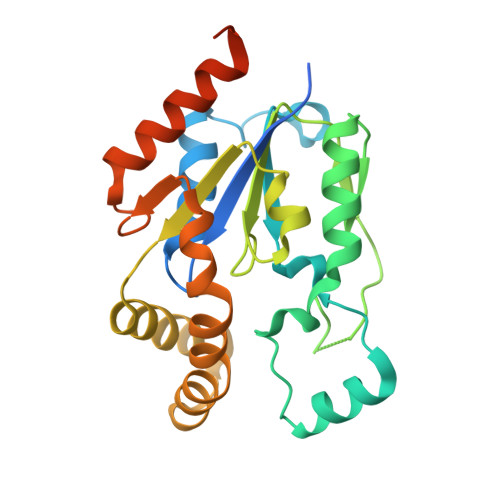 | |
UniProt & NIH Common Fund Data Resources | |||||
PHAROS: Q9BZX2 GTEx: ENSG00000143179 | |||||
Entity Groups | |||||
| Sequence Clusters | 30% Identity50% Identity70% Identity90% Identity95% Identity100% Identity | ||||
| UniProt Group | Q9BZX2 | ||||
Sequence AnnotationsExpand | |||||
Reference Sequence | |||||
| Ligands 3 Unique | |||||
|---|---|---|---|---|---|
| ID | Chains | Name / Formula / InChI Key | 2D Diagram | 3D Interactions | |
| P6D (Subject of Investigation/LOI) Download:Ideal Coordinates CCD File | FA [auth D], MA [auth E], WA [auth H] | 2'-azidocytidine C9 H13 N6 O4 SXKUZFQHWHCRFO-XVFCMESISA-N |  | ||
| PO4 Download:Ideal Coordinates CCD File | GA [auth E] I [auth A] NA [auth F] P [auth B] RA [auth G] | PHOSPHATE ION O4 P NBIIXXVUZAFLBC-UHFFFAOYSA-K |  | ||
| GOL Download:Ideal Coordinates CCD File | AA [auth D] BA [auth D] CA [auth D] DA [auth D] EA [auth D] | GLYCEROL C3 H8 O3 PEDCQBHIVMGVHV-UHFFFAOYSA-N |  | ||
| Length ( Å ) | Angle ( ˚ ) |
|---|---|
| a = 93.73 | α = 90 |
| b = 84.74 | β = 95.36 |
| c = 153.579 | γ = 90 |
| Software Name | Purpose |
|---|---|
| PHENIX | refinement |
| MOSFLM | data reduction |
| Aimless | data scaling |
| PHENIX | phasing |
| Funding Organization | Location | Grant Number |
|---|---|---|
| National Institutes of Health/National Institute of Mental Health (NIH/NIMH) | United States | 1R21MH113062 |


