6POM | pdb_00006pom

Cryo-EM structure of the full-length Bacillus subtilis glyQS T-box riboswitch in complex with tRNA-Gly


Experimental Data Snapshot

  • Method: ELECTRON MICROSCOPY
  • Resolution: 4.90 Å
  • Aggregation State: PARTICLE 
  • Reconstruction Method: SINGLE PARTICLE 

Starting Model: experimental
View more details

wwPDB Validation 3D Report Full Report

Validation slider image for 6POM

This is version 1.4 of the entry. See complete history

Literature

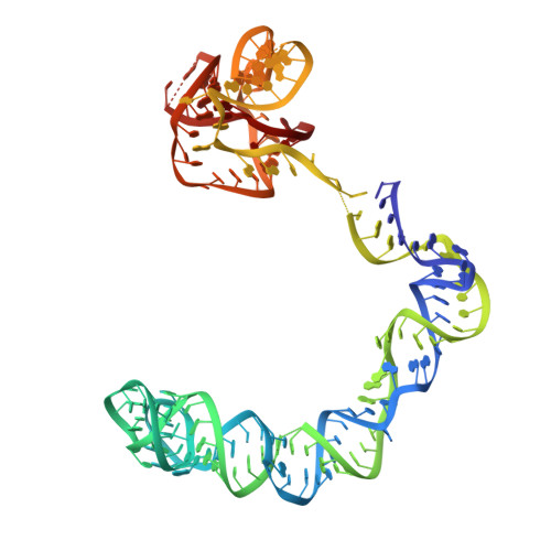
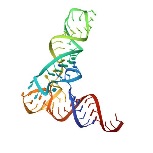
Structural basis of amino acid surveillance by higher-order tRNA-mRNA interactions.

Li, S.Su, Z.Lehmann, J.Stamatopoulou, V.Giarimoglou, N.Henderson, F.E.Fan, L.Pintilie, G.D.Zhang, K.Chen, M.Ludtke, S.J.Wang, Y.X.Stathopoulos, C.Chiu, W.Zhang, J.

(2019) Nat Struct Mol Biol 26: 1094-1105

  • DOI: https://doi.org/10.1038/s41594-019-0326-7
  • Primary Citation Related Structures: 
    6PMO, 6POM

  • PubMed Abstract: 

    Amino acid availability in Gram-positive bacteria is monitored by T-box riboswitches. T-boxes directly bind tRNAs, assess their aminoacylation state, and regulate the transcription or translation of downstream genes to maintain nutritional homeostasis. Here, we report cocrystal and cryo-EM structures of Geobacillus kaustophilus and Bacillus subtilis T-box-tRNA complexes, detailing their multivalent, exquisitely selective interactions. The T-box forms a U-shaped molecular vise that clamps the tRNA, captures its 3' end using an elaborate 'discriminator' structure, and interrogates its aminoacylation state using a steric filter fashioned from a wobble base pair. In the absence of aminoacylation, T-boxes clutch tRNAs and form a continuously stacked central spine, permitting transcriptional readthrough or translation initiation. A modeled aminoacyl disrupts tRNA-T-box stacking, severing the central spine and blocking gene expression. Our data establish a universal mechanism of amino acid sensing on tRNAs and gene regulation by T-box riboswitches and exemplify how higher-order RNA-RNA interactions achieve multivalency and specificity.


  • Organizational Affiliation
    • Laboratory of Molecular Biology, National Institute of Diabetes and Digestive and Kidney Diseases, Bethesda, MD, USA.

Macromolecule Content 

  • Total Structure Weight: 78.89 kDa 
  • Atom Count: 4,937 
  • Modeled Residue Count: 230 
  • Deposited Residue Count: 244 
  • Unique nucleic acid chains: 2

Macromolecules

Find similar nucleic acids by:  (by identity cutoff) 
Entity ID: 1
MoleculeChains LengthOrganismImage
T-box GlyQS leader (155-MER)169Bacillus subtilis
Sequence Annotations
Expand
Reference Sequence
Find similar nucleic acids by:  (by identity cutoff) 
Entity ID: 2
MoleculeChains LengthOrganismImage
tRNAGly (75-MER)75Bacillus subtilis
Sequence Annotations
Expand
Reference Sequence

Experimental Data & Validation

Experimental Data

  • Method: ELECTRON MICROSCOPY
  • Resolution: 4.90 Å
  • Aggregation State: PARTICLE 
  • Reconstruction Method: SINGLE PARTICLE 
EM Software:
TaskSoftware PackageVersion
RECONSTRUCTIONRELION3.0-beta
RECONSTRUCTIONcryoSPARC2.2
MODEL REFINEMENTPHENIX1.15.2

Structure Validation

View Full Validation Report


Entry History 

& Funding Information

Deposition Data


Funding OrganizationLocationGrant Number
National Institutes of Health/National Institute of General Medical Sciences (NIH/NIGMS)United StatesP50GM103297

Revision History  (Full details and data files)

  • Version 1.0: 2019-11-20
    Type: Initial release
  • Version 1.1: 2019-12-04
    Changes: Database references
  • Version 1.2: 2019-12-18
    Changes: Database references
  • Version 1.3: 2020-01-08
    Changes: Author supporting evidence
  • Version 1.4: 2024-03-20
    Changes: Data collection, Database references, Refinement description