






 Download Mendeley
High resolution X-ray structure of glucagon and selected stereo-inversed analogs in novel crystallographic packing arrangement.
Mroz, P.A., Gonzalez-Gutierrez, G., DiMarchi, R.D.To be published.
Experimental Data Snapshot
wwPDB Validation 3D Report Full Report

Entity ID: 1 | |||||
|---|---|---|---|---|---|
| Molecule | Chains | Sequence Length | Organism | Details | Image |
| D-glucagon D-BrPhe 6,22 | 26 | Homo sapiens | Mutation(s): 0 | 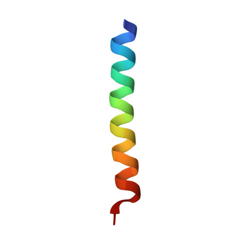 | |
Sequence AnnotationsExpand | |||||
Reference Sequence | |||||
| Modified Residues 14 Unique | |||||
|---|---|---|---|---|---|
| ID | Chains | Type | Formula | 2D Diagram | Parent |
| BF7 Query on BF7 | A | D-PEPTIDE LINKING | C9 H10 Br N O2 | 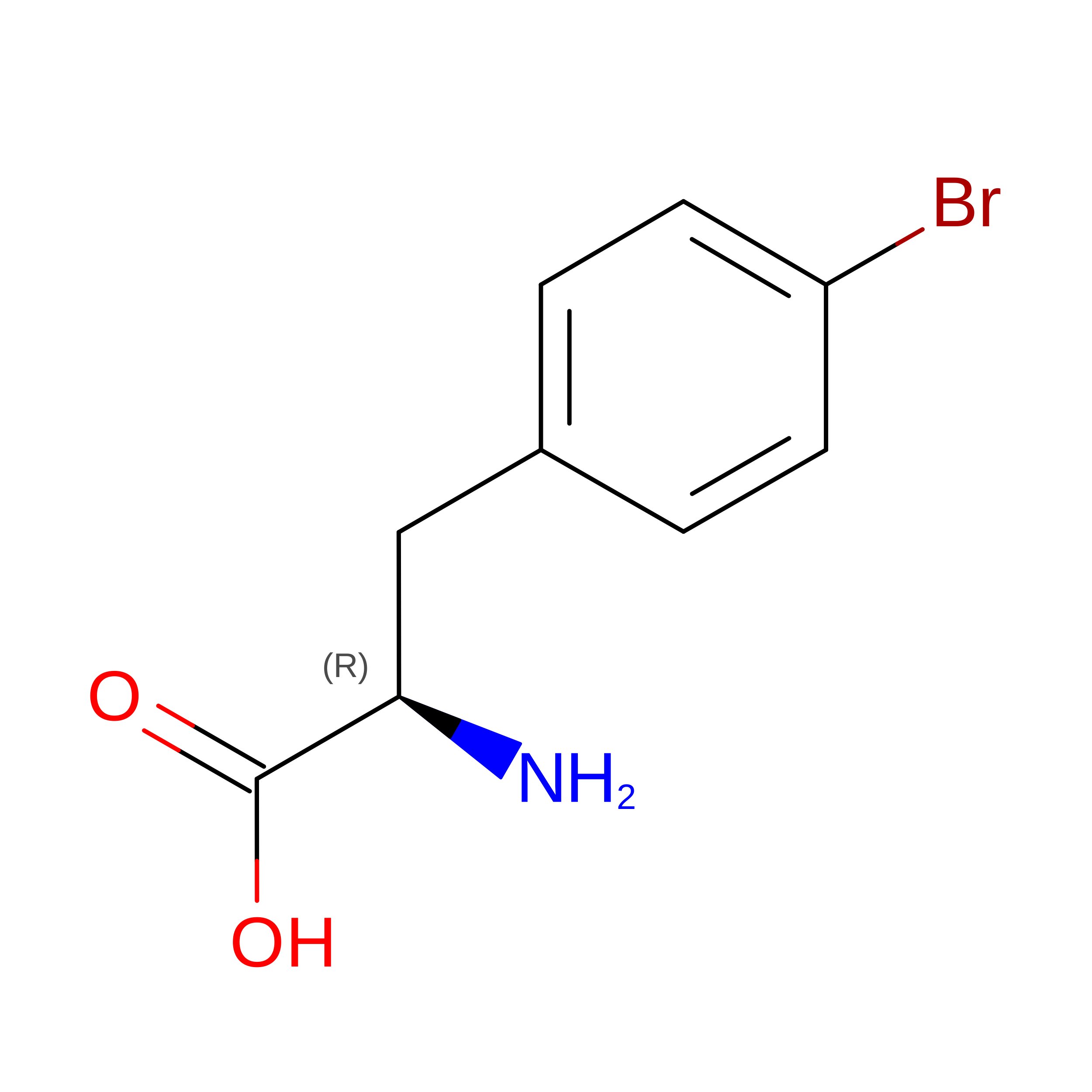 | -- |
| DAL Query on DAL | A | D-PEPTIDE LINKING | C3 H7 N O2 | 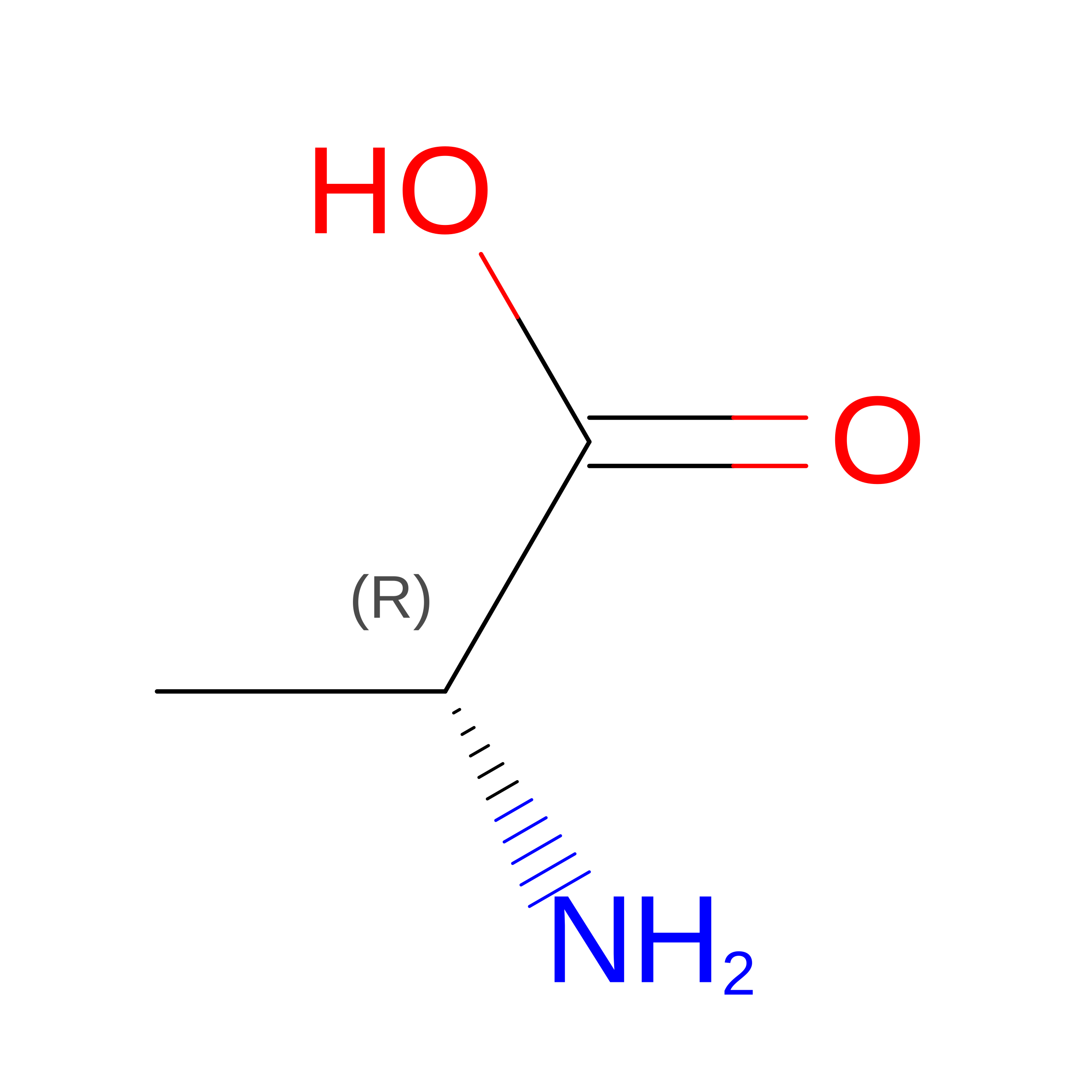 | -- |
| DAR Query on DAR | A | D-PEPTIDE LINKING | C6 H15 N4 O2 | 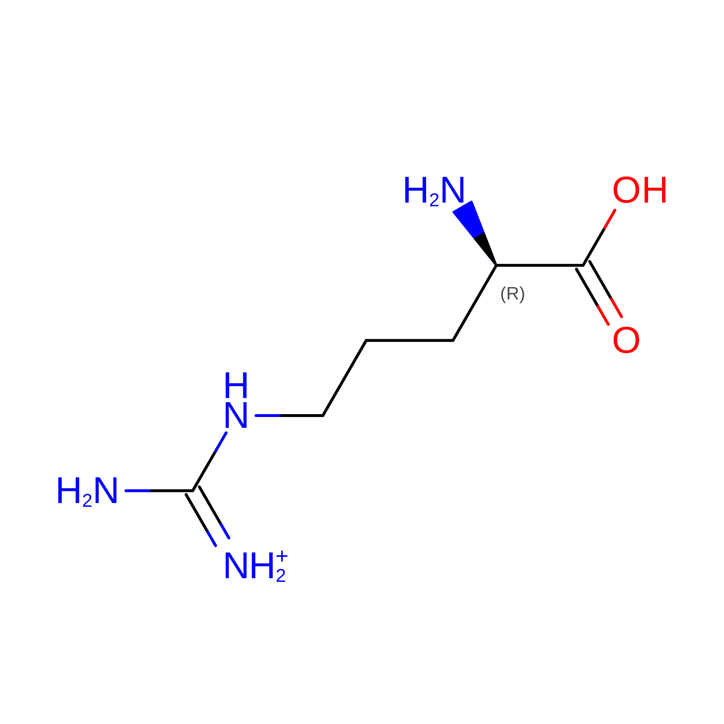 | -- |
| DAS Query on DAS | A | D-PEPTIDE LINKING | C4 H7 N O4 | 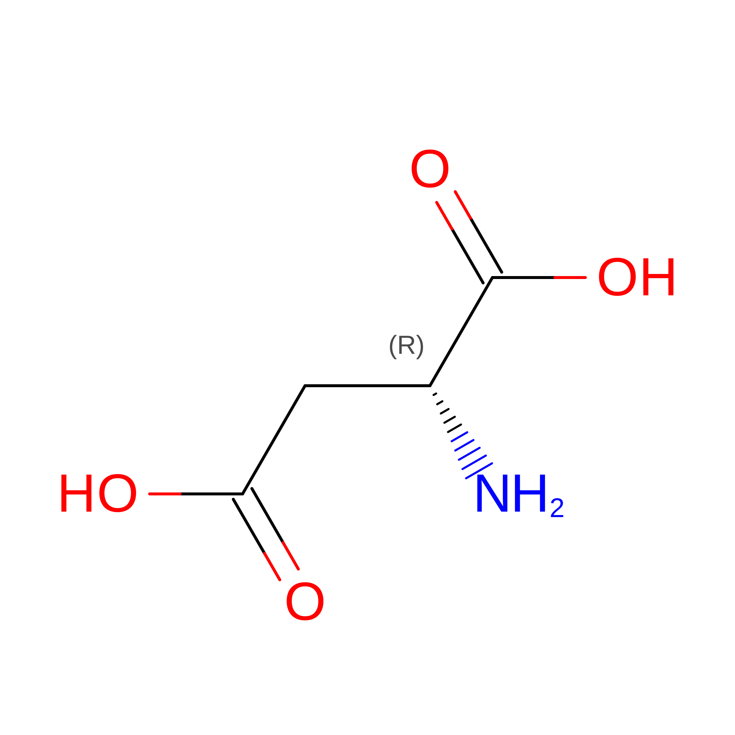 | -- |
| DGN Query on DGN | A | D-PEPTIDE LINKING | C5 H10 N2 O3 | 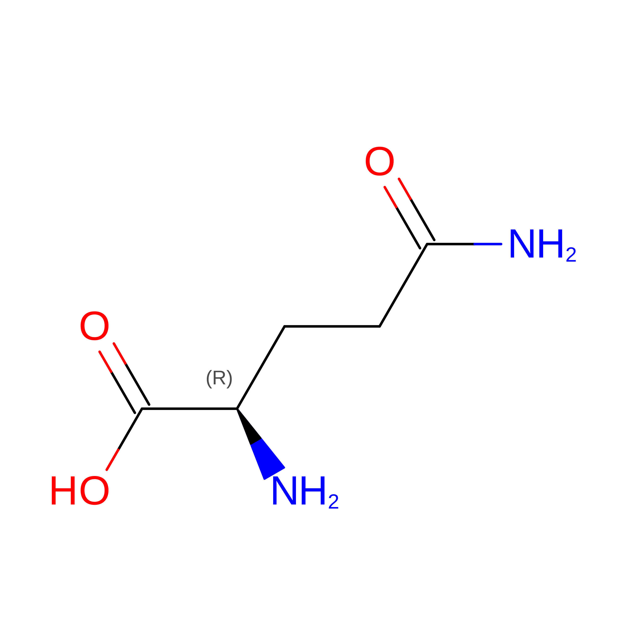 | -- |
| DLE Query on DLE | A | D-PEPTIDE LINKING | C6 H13 N O2 | 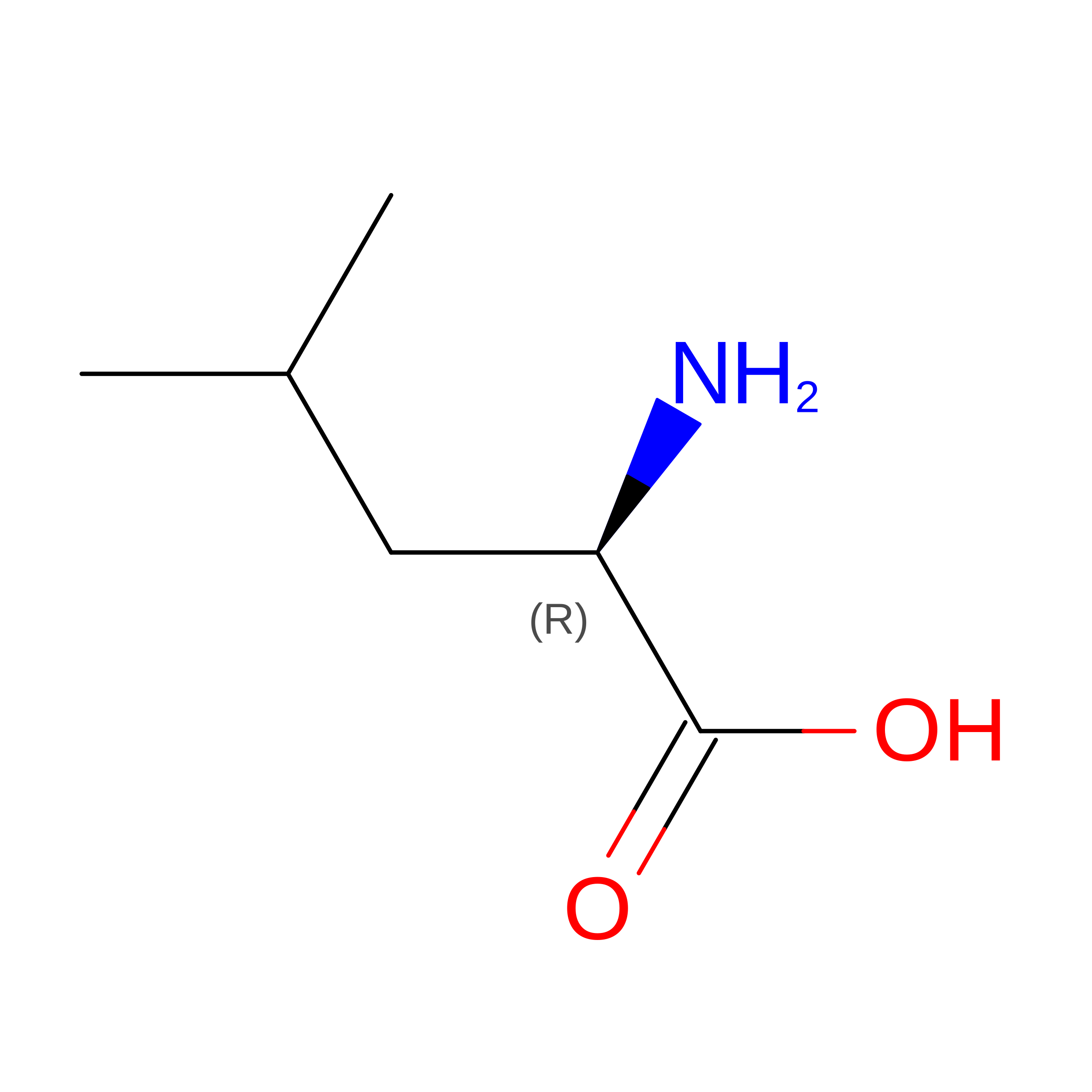 | -- |
| DLY Query on DLY | A | D-PEPTIDE LINKING | C6 H14 N2 O2 | 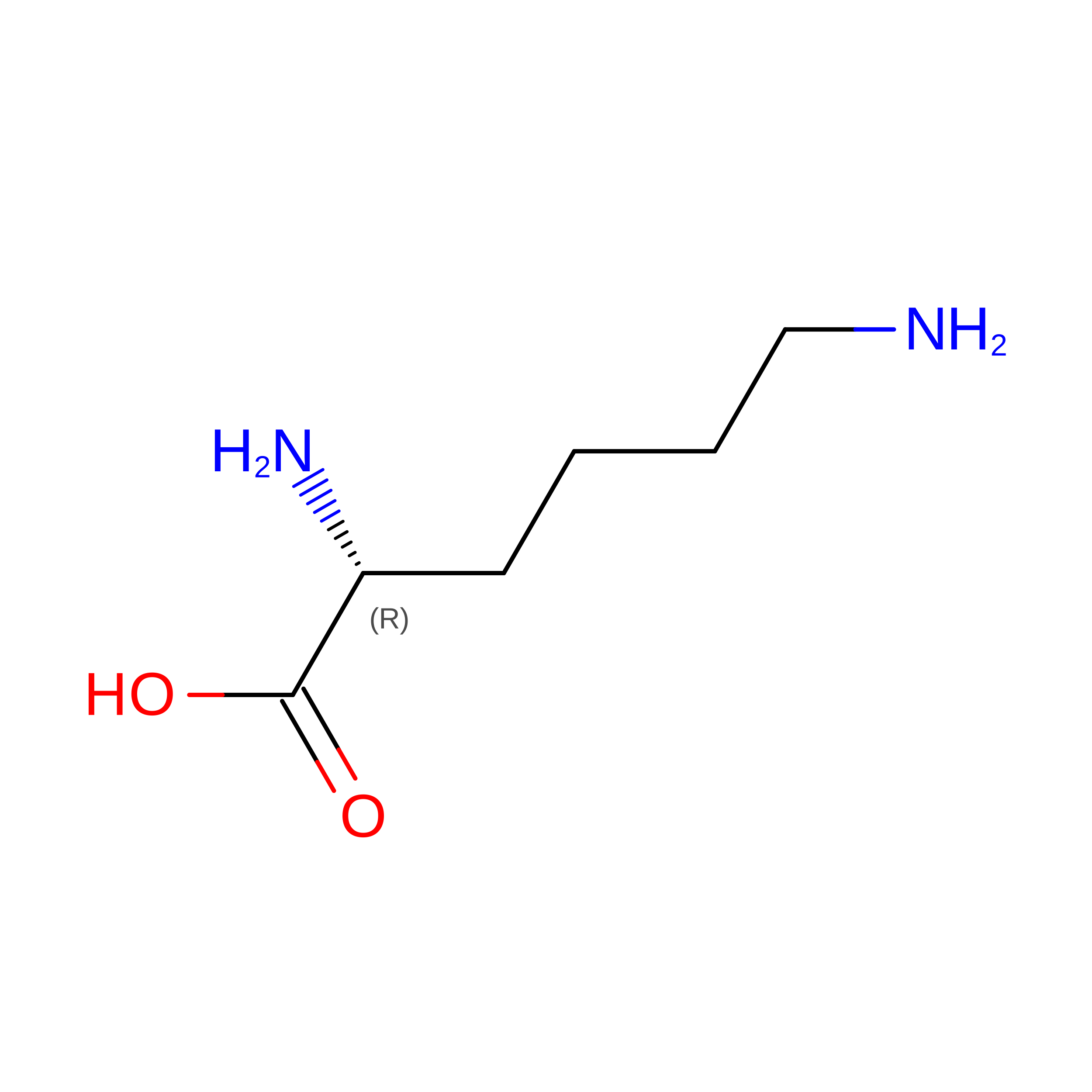 | -- |
| DSG Query on DSG | A | D-PEPTIDE LINKING | C4 H8 N2 O3 | 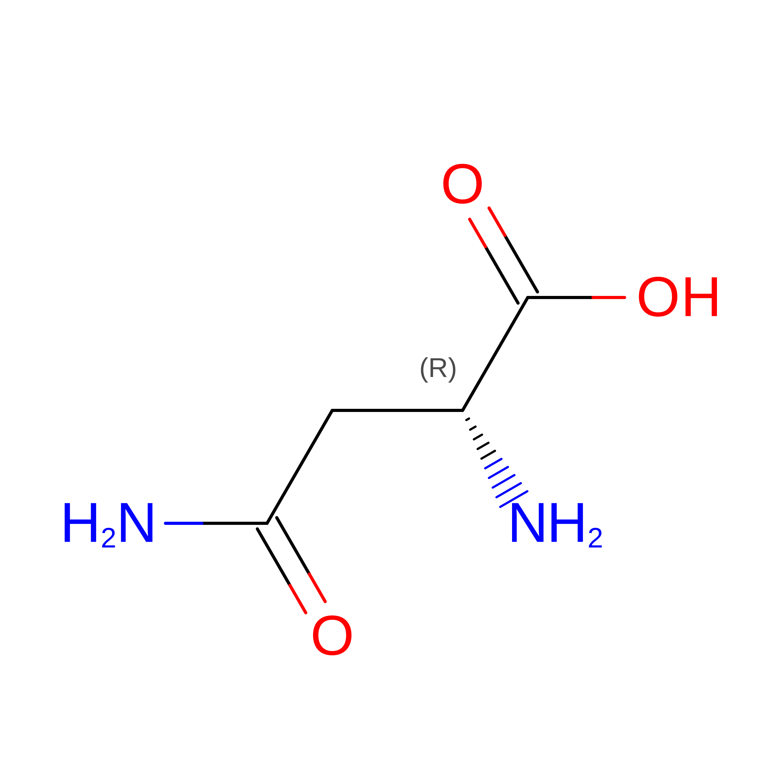 | -- |
| DSN Query on DSN | A | D-PEPTIDE LINKING | C3 H7 N O3 | 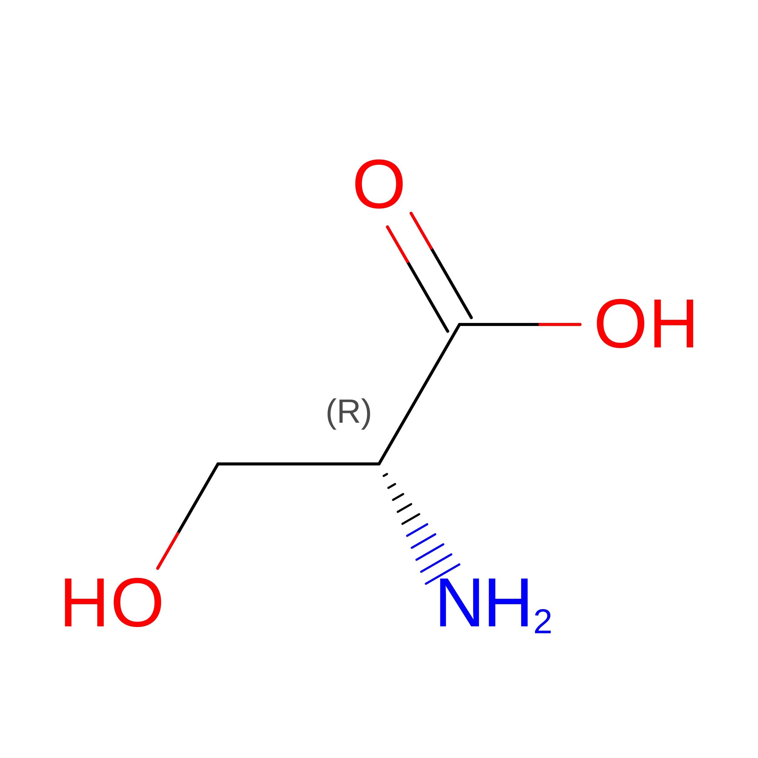 | -- |
| DTH Query on DTH | A | D-PEPTIDE LINKING | C4 H9 N O3 | 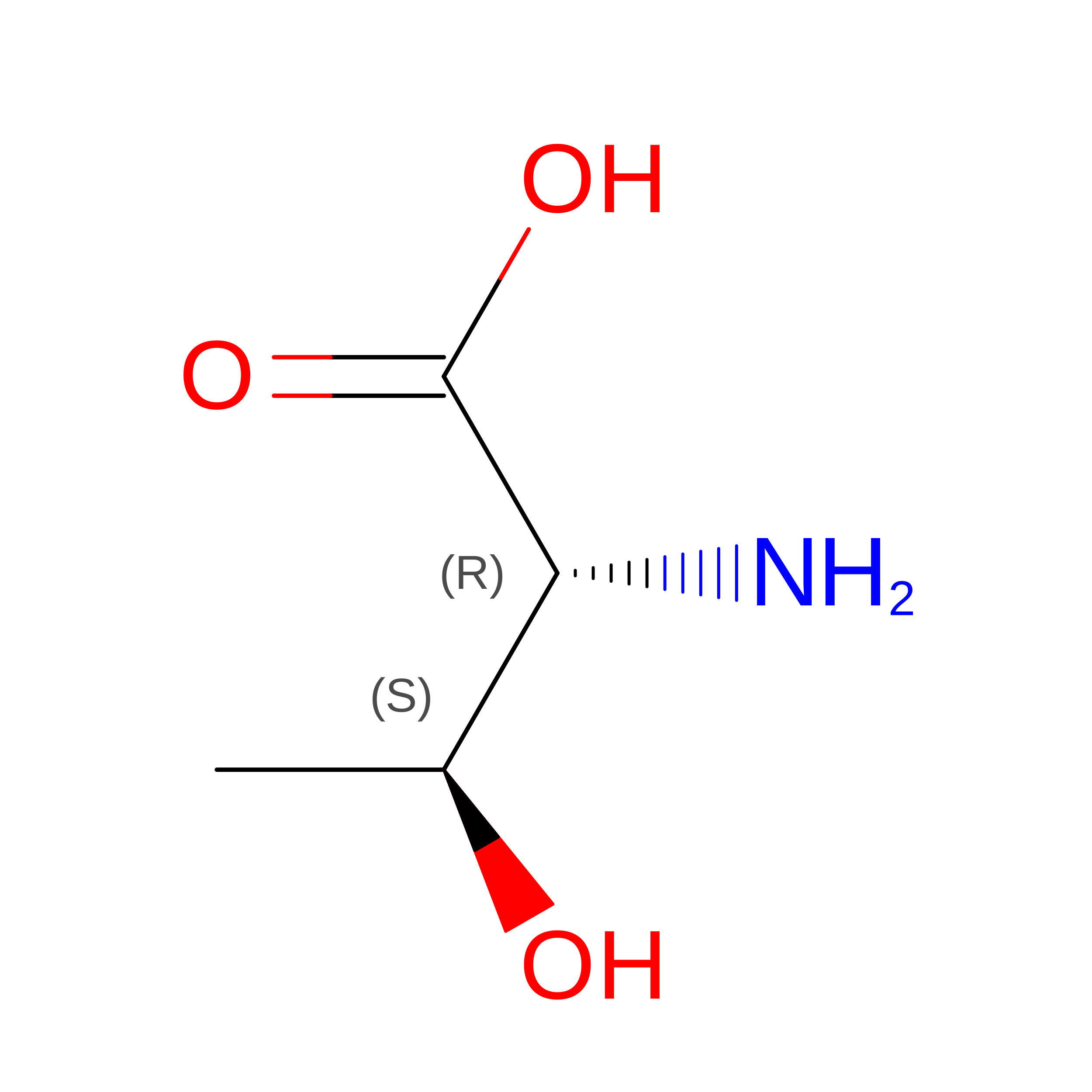 | -- |
| DTR Query on DTR | A | D-PEPTIDE LINKING | C11 H12 N2 O2 | 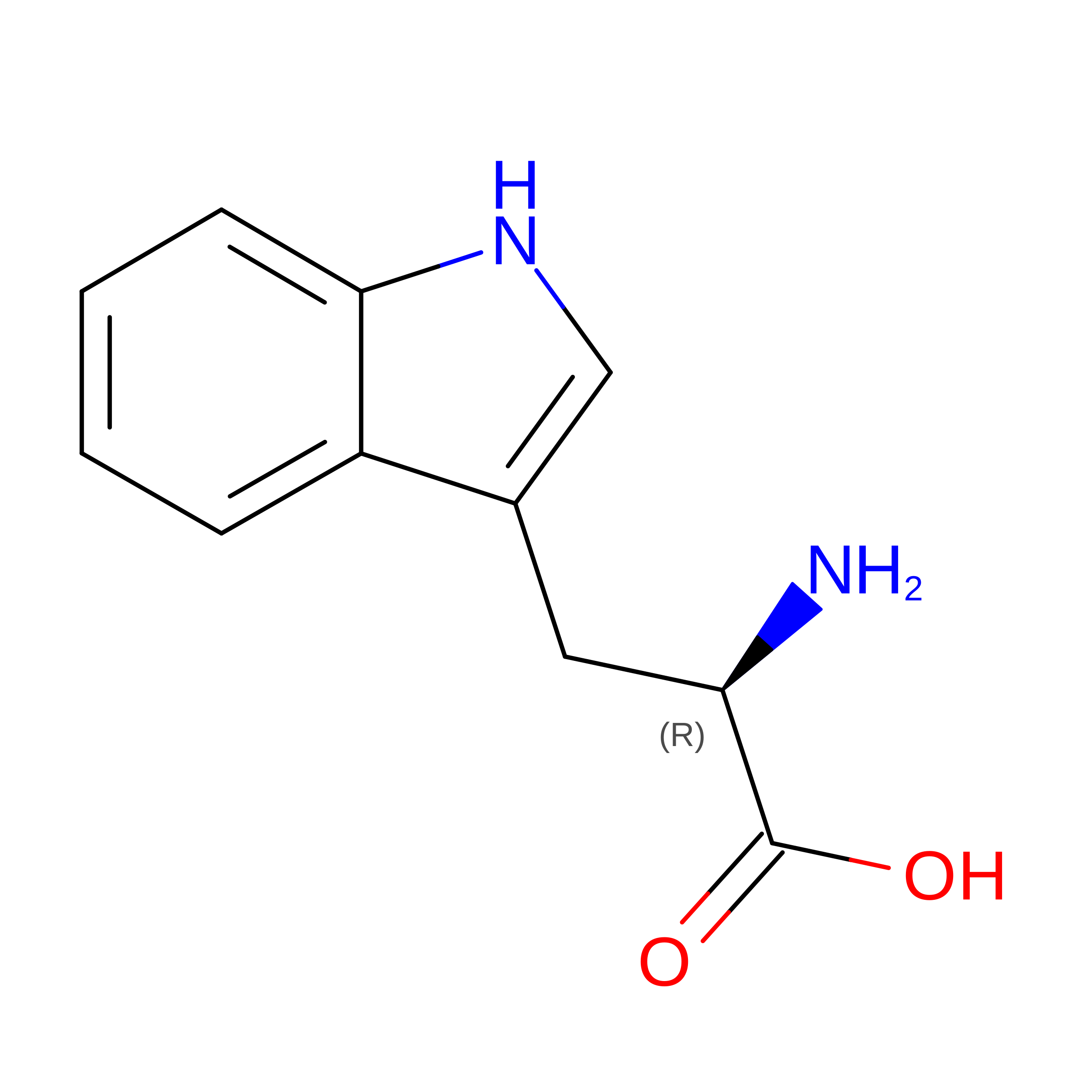 | -- |
| DTY Query on DTY | A | D-PEPTIDE LINKING | C9 H11 N O3 | 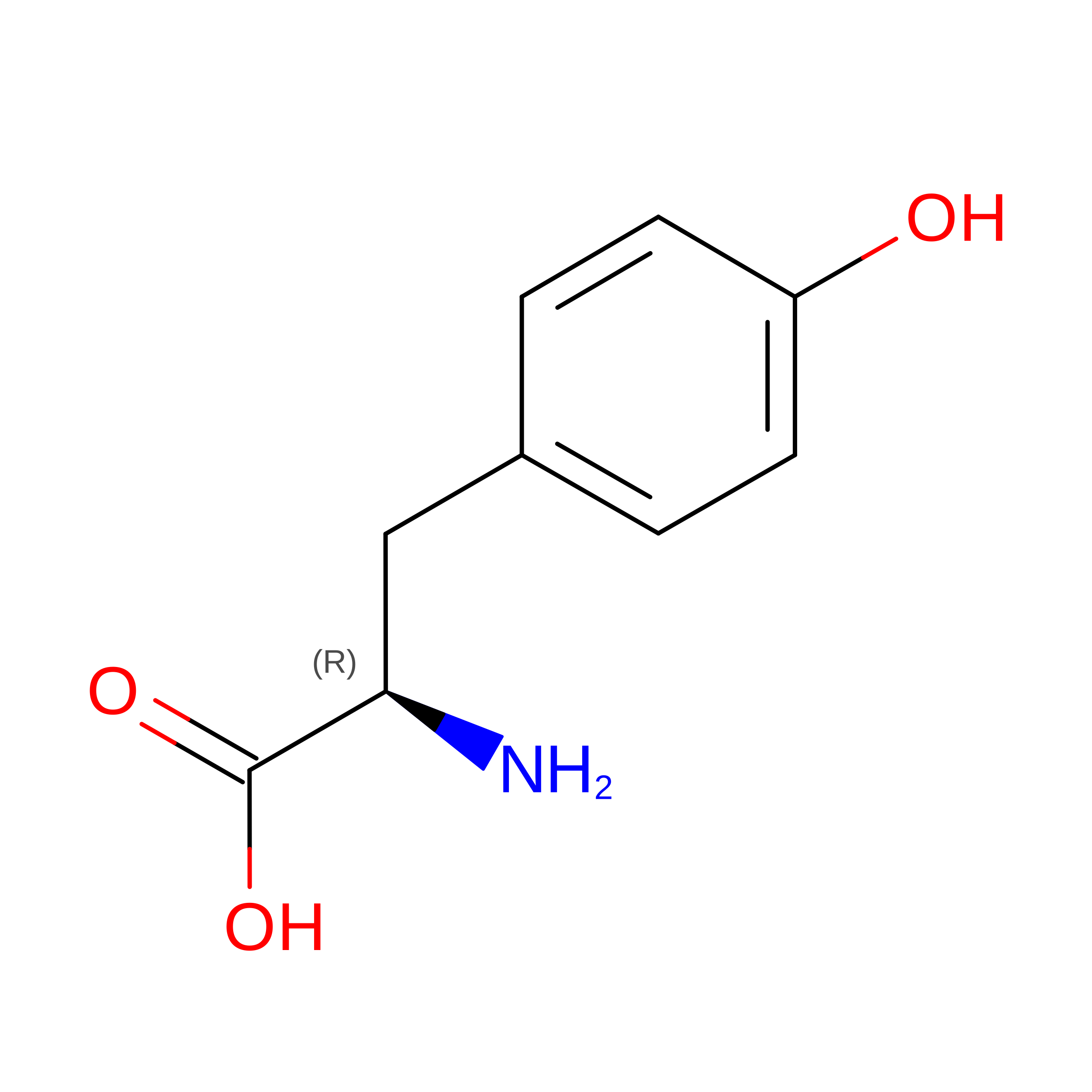 | -- |
| DVA Query on DVA | A | D-PEPTIDE LINKING | C5 H11 N O2 | 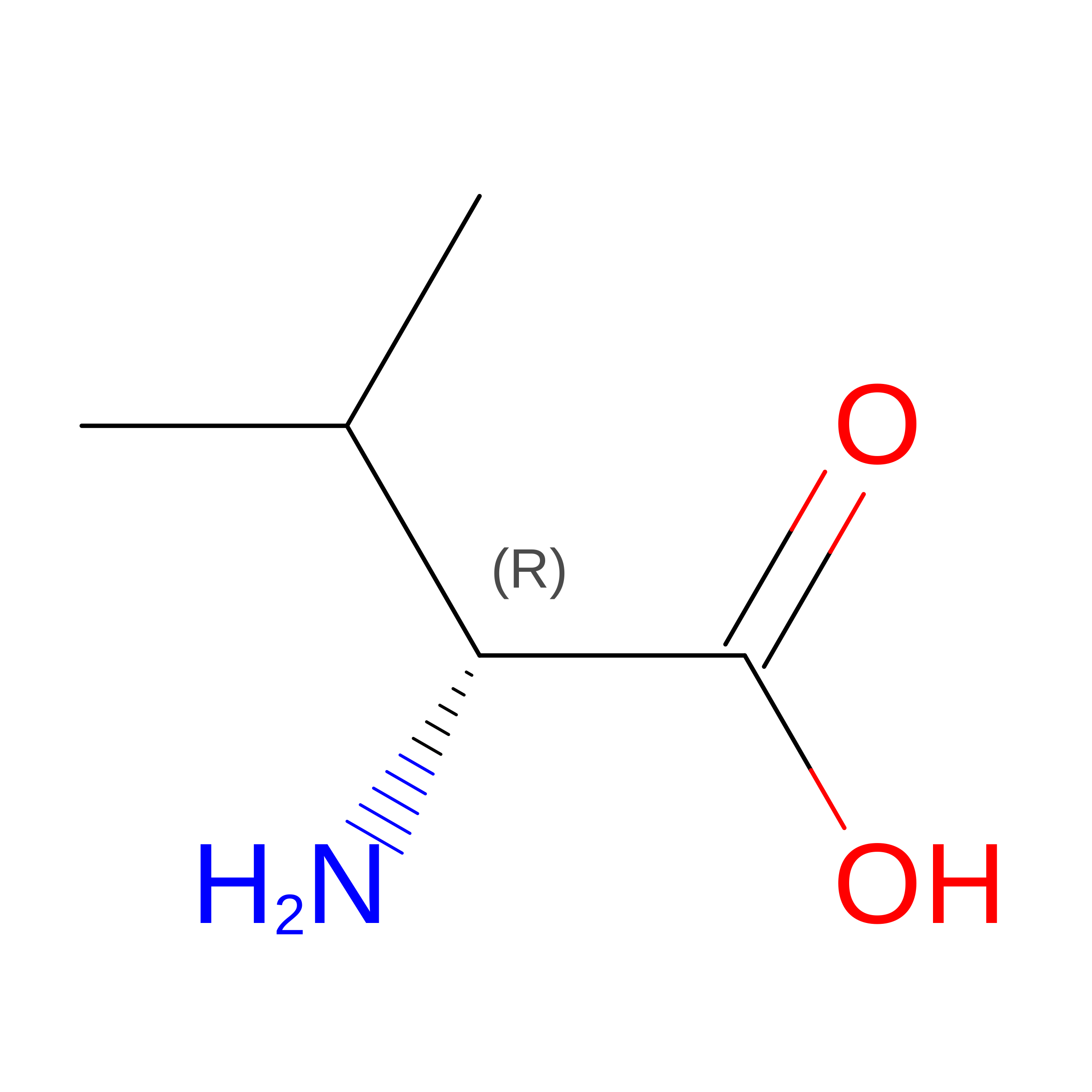 | -- |
| MED Query on MED | A | D-PEPTIDE LINKING | C5 H11 N O2 S | 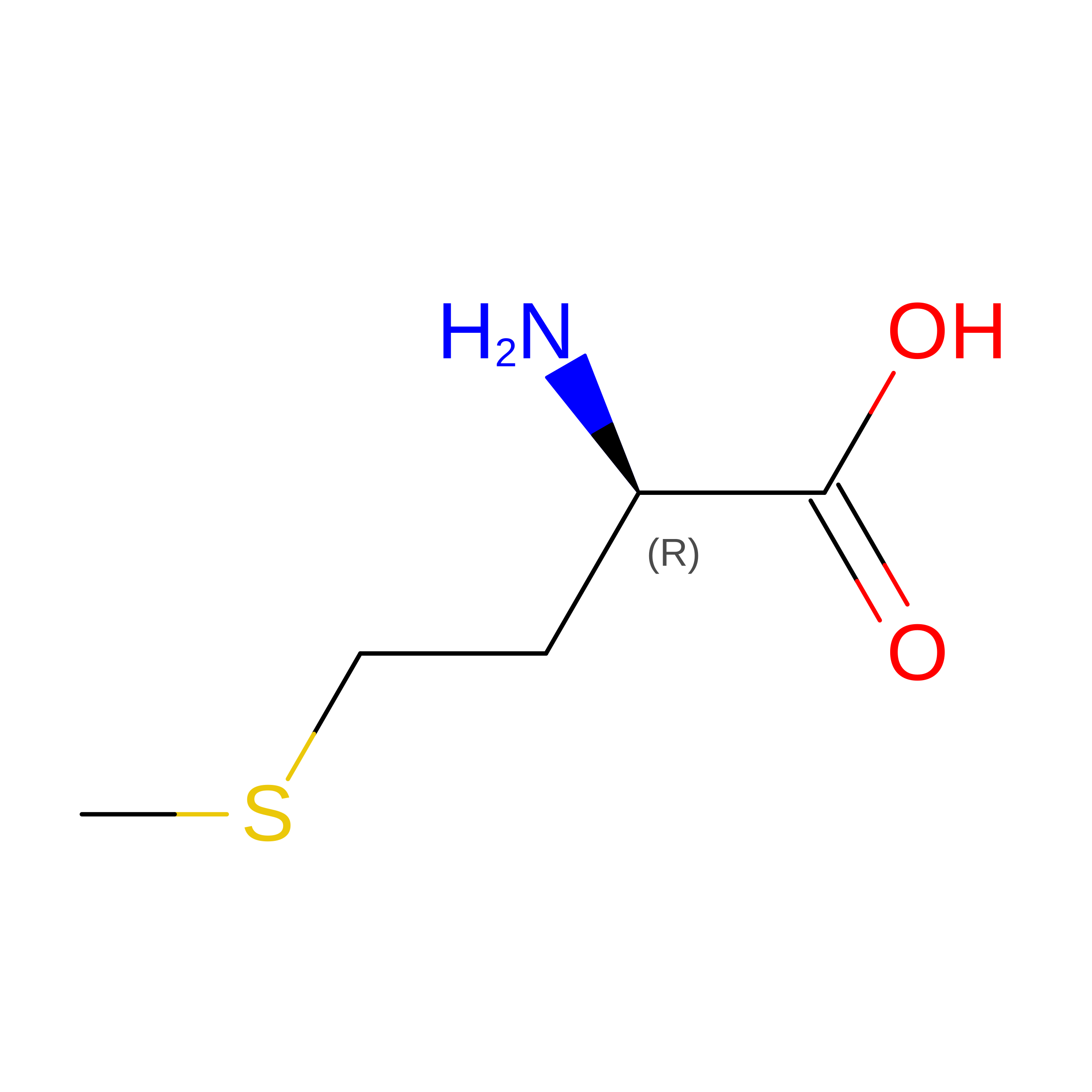 | -- |
| Length ( Å ) | Angle ( ˚ ) |
|---|---|
| a = 41.77 | α = 90 |
| b = 41.77 | β = 90 |
| c = 30.439 | γ = 90 |
| Software Name | Purpose |
|---|---|
| PHENIX | refinement |
| XDS | data reduction |
| Aimless | data scaling |
| PHASER | phasing |
| Funding Organization | Location | Grant Number |
|---|---|---|
| Other government | United States | Indiana University Internal grant |








