6PGW | pdb_00006pgw

Crystal structure of zebrafish Protocadherin-19 EC3-6


Experimental Data Snapshot

  • Method: X-RAY DIFFRACTION
  • Resolution: 3.00 Å
  • R-Value Free: 
    0.298 (Depositor), 0.300 (DCC) 
  • R-Value Work: 
    0.215 (Depositor), 0.218 (DCC) 
  • R-Value Observed: 
    0.219 (Depositor) 

Starting Model: experimental
View more details

wwPDB Validation 3D Report Full Report

Validation slider image for 6PGW

This is version 1.2 of the entry. See complete history

Literature

A complete Protocadherin-19 ectodomain model for evaluating epilepsy-causing mutations and potential protein interaction sites.

Hudson, J.D.Tamilselvan, E.Sotomayor, M.Cooper, S.R.

(2021) Structure 29: 1128-1143.e4

  • DOI: https://doi.org/10.1016/j.str.2021.07.006
  • Primary Citation Related Structures: 
    6PGW

  • PubMed Abstract: 

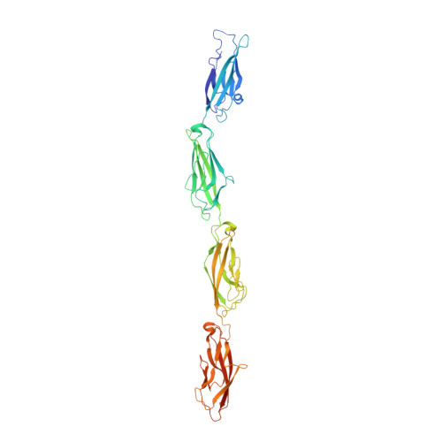
    Cadherin superfamily members play a critical role in differential adhesion during neurodevelopment, and their disruption has been linked to several neurodevelopmental disorders. Mutations in protocadherin-19 (PCDH19), a member of the δ-protocadherin subfamily of cadherins, cause a unique form of epilepsy called PCDH19 clustering epilepsy. While PCDH19 and other non-clustered δ-protocadherins form multimers with other members of the cadherin superfamily to alter adhesiveness, the specific protein surfaces responsible for these interactions are unknown. Only portions of the PCDH19 extracellular domain structure had been solved previously. Here, we present a structure of the missing segment from zebrafish Protocadherin-19 (Pcdh19) and create a complete ectodomain model. This model shows the structural environment for 97% of disease-causing missense mutations and reveals two potential surfaces for intermolecular interactions that could modify Pcdh19's adhesive strength and specificity.


  • Organizational Affiliation
    • Department of Science and Mathematics, Cedarville University, 251 N. Main Street, Cedarville, OH 45314, USA.

Macromolecule Content 

  • Total Structure Weight: 49.22 kDa 
  • Atom Count: 3,323 
  • Modeled Residue Count: 427 
  • Deposited Residue Count: 443 
  • Unique protein chains: 1

Macromolecules

Find similar proteins by:|  3D Structure
Entity ID: 1
MoleculeChains  Sequence LengthOrganismDetailsImage
Protocadherin-19443Danio rerioMutation(s): 0 
Gene Names: pcdh19wu:fc83e05
UniProt
Find proteins for F8W3X3 (Danio rerio)
Explore F8W3X3 
Go to UniProtKB:  F8W3X3
Entity Groups
Sequence Clusters30% Identity50% Identity70% Identity90% Identity95% Identity100% Identity
UniProt GroupF8W3X3
Sequence Annotations
Expand
Reference Sequence

Small Molecules

Ligands 2 Unique
IDChains Name / Formula / InChI Key2D Diagram3D Interactions
CA

Query on CA


Download:Ideal Coordinates CCD File
B [auth A]
C [auth A]
D [auth A]
E [auth A]
F [auth A]
B [auth A],
C [auth A],
D [auth A],
E [auth A],
F [auth A],
G [auth A],
H [auth A],
I [auth A],
J [auth A]
CALCIUM ION
Ca
BHPQYMZQTOCNFJ-UHFFFAOYSA-N
NA

Query on NA


Download:Ideal Coordinates CCD File
K [auth A]SODIUM ION
Na
FKNQFGJONOIPTF-UHFFFAOYSA-N

Experimental Data & Validation

Experimental Data

  • Method: X-RAY DIFFRACTION
  • Resolution: 3.00 Å
  • R-Value Free:  0.298 (Depositor), 0.300 (DCC) 
  • R-Value Work:  0.215 (Depositor), 0.218 (DCC) 
  • R-Value Observed: 0.219 (Depositor) 
Space Group: P 1 21 1
Unit Cell:
Length ( Å )Angle ( ˚ )
a = 69.92α = 90
b = 58.896β = 117.97
c = 70.956γ = 90
Software Package:
Software NamePurpose
REFMACrefinement
HKL-2000data reduction
HKL-2000data scaling
PHASERphasing

Structure Validation

View Full Validation Report


Entry History 

Deposition Data

Revision History  (Full details and data files)

  • Version 1.0: 2020-12-30
    Type: Initial release
  • Version 1.1: 2021-12-01
    Changes: Database references
  • Version 1.2: 2023-10-11
    Changes: Data collection, Refinement description