6OVM | pdb_00006ovm

Crystal Structure of the Pseudomonas capeferrum Anti-sigma Regulator PupR C-terminal Cell-surface Signaling Domain in Complex with the Outer Membrane Transporter PupB N-terminal Signaling Domain (SeMet)


Experimental Data Snapshot

  • Method: X-RAY DIFFRACTION
  • Resolution: 1.60 Å
  • R-Value Free: 
    0.184 (Depositor), 0.189 (DCC) 
  • R-Value Work: 
    0.153 (Depositor), 0.165 (DCC) 
  • R-Value Observed: 
    0.154 (Depositor) 

wwPDB Validation 3D Report Full Report

Validation slider image for 6OVM

This is version 1.4 of the entry. See complete history

Literature

Structural basis of cell-surface signaling by a conserved sigma regulator in Gram-negative bacteria.

Jensen, J.L.Jernberg, B.D.Sinha, S.C.Colbert, C.L.

(2020) J Biological Chem 295: 5795-5806

  • DOI: https://doi.org/10.1074/jbc.RA119.010697
  • Primary Citation Related Structures: 
    6OVK, 6OVM

  • PubMed Abstract: 

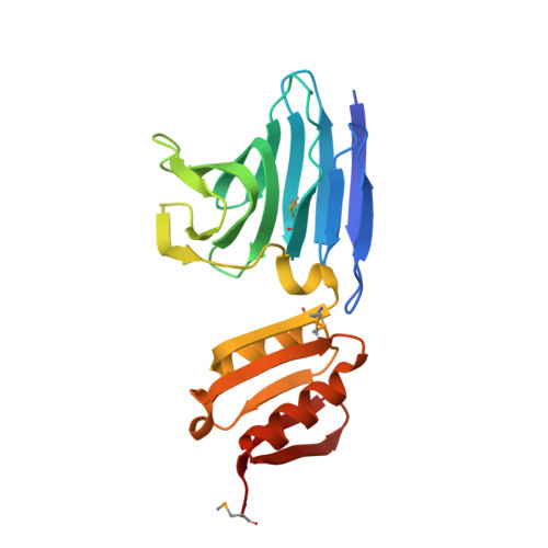
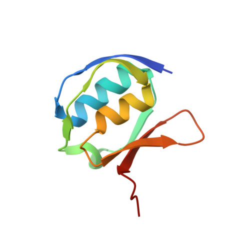
    Cell-surface signaling (CSS) in Gram-negative bacteria involves highly conserved regulatory pathways that optimize gene expression by transducing extracellular environmental signals to the cytoplasm via inner-membrane sigma regulators. The molecular details of ferric siderophore-mediated activation of the iron import machinery through a sigma regulator are unclear. Here, we present the 1.56 Å resolution structure of the periplasmic complex of the C-terminal CSS domain (CCSSD) of PupR, the sigma regulator in the Pseudomonas capeferrum pseudobactin BN7/8 transport system, and the N-terminal signaling domain (NTSD) of PupB, an outer-membrane TonB-dependent transducer. The structure revealed that the CCSSD consists of two subdomains: a juxta-membrane subdomain, which has a novel all-β-fold, followed by a secretin/TonB, short N-terminal subdomain at the C terminus of the CCSSD, a previously unobserved topological arrangement of this domain. Using affinity pulldown assays, isothermal titration calorimetry, and thermal denaturation CD spectroscopy, we show that both subdomains are required for binding the NTSD with micromolar affinity and that NTSD binding improves CCSSD stability. Our findings prompt us to present a revised model of CSS wherein the CCSSD:NTSD complex forms prior to ferric-siderophore binding. Upon siderophore binding, conformational changes in the CCSSD enable regulated intramembrane proteolysis of the sigma regulator, ultimately resulting in transcriptional regulation.


  • Organizational Affiliation
    • Department of Chemistry and Biochemistry, North Dakota State University, Fargo, North Dakota 58108.

Macromolecule Content 

  • Total Structure Weight: 32.75 kDa 
  • Atom Count: 2,789 
  • Modeled Residue Count: 293 
  • Deposited Residue Count: 301 
  • Unique protein chains: 2

Macromolecules

Find similar proteins by:|  3D Structure
Entity ID: 1
MoleculeChains  Sequence LengthOrganismDetailsImage
Siderophore-interacting proteinA [auth R]219Pseudomonas capeferrumMutation(s): 0 
Gene Names: PC358_08100pupR
Find similar proteins by:|  3D Structure
Entity ID: 2
MoleculeChains  Sequence LengthOrganismDetailsImage
Ferric-pseudobactin BN7/BN8 receptor82Pseudomonas capeferrumMutation(s): 0 
Gene Names: PC358_08105pupB

Small Molecules

Modified Residues  1 Unique
IDChains TypeFormula2D DiagramParent
MSE
Query on MSE
A [auth R]L-PEPTIDE LINKINGC5 H11 N O2 SeMET

Experimental Data & Validation

Experimental Data

  • Method: X-RAY DIFFRACTION
  • Resolution: 1.60 Å
  • R-Value Free:  0.184 (Depositor), 0.189 (DCC) 
  • R-Value Work:  0.153 (Depositor), 0.165 (DCC) 
  • R-Value Observed: 0.154 (Depositor) 
Space Group: P 21 21 21
Unit Cell:
Length ( Å )Angle ( ˚ )
a = 43.574α = 90
b = 44.714β = 90
c = 141.282γ = 90
Software Package:
Software NamePurpose
PHENIXrefinement
HKL-2000data reduction
SCALAdata scaling
PHASERphasing

Structure Validation

View Full Validation Report


Entry History 

& Funding Information

Deposition Data


Funding OrganizationLocationGrant Number
National Institutes of Health/National Institute of General Medical Sciences (NIH/NIGMS)United States1R15GM113227-01

Revision History  (Full details and data files)

  • Version 1.0: 2020-03-04
    Type: Initial release
  • Version 1.1: 2020-03-11
    Changes: Database references
  • Version 1.2: 2020-05-06
    Changes: Database references
  • Version 1.3: 2020-05-13
    Changes: Database references
  • Version 1.4: 2024-10-16
    Changes: Advisory, Data collection, Database references, Structure summary