6OTN

Crystal Structure of an N-terminal Fragment of Cancer Associated Tropomyosin 3.1 (Tpm3.1)


Experimental Data Snapshot

  • Method: X-RAY DIFFRACTION
  • Resolution: 2.40 Å
  • R-Value Free: 0.266 
  • R-Value Work: 0.235 
  • R-Value Observed: 0.238 

wwPDB Validation   3D Report Full Report


This is version 1.2 of the entry. See complete history


Literature

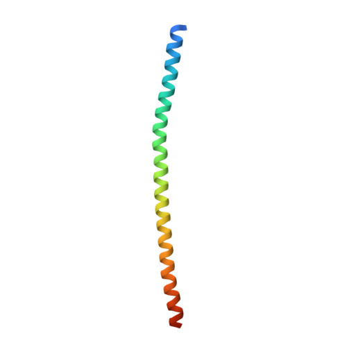
Molecular integration of the anti-tropomyosin compound ATM-3507 into the coiled coil overlap region of the cancer-associated Tpm3.1.

Janco, M.Rynkiewicz, M.J.Li, L.Hook, J.Eiffe, E.Ghosh, A.Bocking, T.Lehman, W.J.Hardeman, E.C.Gunning, P.W.

(2019) Sci Rep 9: 11262-11262

  • DOI: https://doi.org/10.1038/s41598-019-47592-9
  • Primary Citation of Related Structures:  
    6OTN

  • PubMed Abstract: 

    Tropomyosins (Tpm) determine the functional capacity of actin filaments in an isoform-specific manner. The primary isoform in cancer cells is Tpm3.1 and compounds that target Tpm3.1 show promising results as anti-cancer agents both in vivo and in vitro. We have determined the molecular mechanism of interaction of the lead compound ATM-3507 with Tpm3.1-containing actin filaments. When present during co-polymerization of Tpm3.1 with actin, 3 H-ATM-3507 is incorporated into the filaments and saturates at approximately one molecule per Tpm3.1 dimer and with an apparent binding affinity of approximately 2 µM. In contrast, 3 H-ATM-3507 is poorly incorporated into preformed Tpm3.1/actin co-polymers. CD spectroscopy and thermal melts using Tpm3.1 peptides containing the C-terminus, the N-terminus, and a combination of the two forming the overlap junction at the interface of adjacent Tpm3.1 dimers, show that ATM-3507 shifts the melting temperature of the C-terminus and the overlap junction, but not the N-terminus. Molecular dynamic simulation (MDS) analysis predicts that ATM-3507 integrates into the 4-helix coiled coil overlap junction and in doing so, likely changes the lateral movement of Tpm3.1 across the actin surface resulting in an alteration of filament interactions with actin binding proteins and myosin motors, consistent with the cellular impact of ATM-3507.


  • Organizational Affiliation

    School of Medical Sciences, University of New South Wales Sydney, Sydney, NSW, 2052, Australia.


Macromolecules
Find similar proteins by:  (by identity cutoff)  |  3D Structure
Entity ID: 1
MoleculeChains Sequence LengthOrganismDetailsImage
Tropomyosin alpha-3 chain
A, B, C, D
81Homo sapiensMutation(s): 1 
Gene Names: TPM3
UniProt & NIH Common Fund Data Resources
Find proteins for P06753 (Homo sapiens)
Explore P06753 
Go to UniProtKB:  P06753
PHAROS:  P06753
GTEx:  ENSG00000143549 
Entity Groups  
Sequence Clusters30% Identity50% Identity70% Identity90% Identity95% Identity100% Identity
UniProt GroupP06753
Sequence Annotations
Expand
  • Reference Sequence
Small Molecules
Ligands 1 Unique
IDChains Name / Formula / InChI Key2D Diagram3D Interactions
SO4
Query on SO4

Download Ideal Coordinates CCD File 
E [auth A]SULFATE ION
O4 S
QAOWNCQODCNURD-UHFFFAOYSA-L
Experimental Data & Validation

Experimental Data

  • Method: X-RAY DIFFRACTION
  • Resolution: 2.40 Å
  • R-Value Free: 0.266 
  • R-Value Work: 0.235 
  • R-Value Observed: 0.238 
  • Space Group: P 43 21 2
Unit Cell:
Length ( Å )Angle ( ˚ )
a = 97.201α = 90
b = 97.201β = 90
c = 93.1γ = 90
Software Package:
Software NamePurpose
XDSdata reduction
Aimlessdata scaling
PHASERphasing
PHENIXrefinement
PDB_EXTRACTdata extraction

Structure Validation

View Full Validation Report


Entry History & Funding Information

Deposition Data


Funding OrganizationLocationGrant Number
National Health and Medical Research Council (NHMRC, Australia)AustraliaAPP1100202
National Health and Medical Research Council (NHMRC, Australia)AustraliaAPP1079866

Revision History  (Full details and data files)

  • Version 1.0: 2019-08-14
    Type: Initial release
  • Version 1.1: 2020-01-08
    Changes: Author supporting evidence
  • Version 1.2: 2023-10-11
    Changes: Data collection, Database references, Refinement description