6OM4 | pdb_00006om4

The structure of Microcin C7 biosynthetic enzyme MccB in complex with N-formylated MccA


Experimental Data Snapshot

  • Method: X-RAY DIFFRACTION
  • Resolution: 1.70 Å
  • R-Value Free: 
    0.199 (Depositor), 0.206 (DCC) 
  • R-Value Work: 
    0.155 (Depositor), 0.165 (DCC) 
  • R-Value Observed: 
    0.157 (Depositor) 

Starting Model: experimental
View more details

wwPDB Validation 3D Report Full Report

Validation slider image for 6OM4

Ligand Structure Quality Assessment 


This is version 1.3 of the entry. See complete history

Literature

Biosynthesis of the RiPP trojan horse nucleotide antibiotic microcin C is directed by theN-formyl of the peptide precursor.

Dong, S.H.Kulikovsky, A.Zukher, I.Estrada, P.Dubiley, S.Severinov, K.Nair, S.K.

(2019) Chem Sci 10: 2391-2395

  • DOI: https://doi.org/10.1039/c8sc03173h
  • Primary Citation Related Structures: 
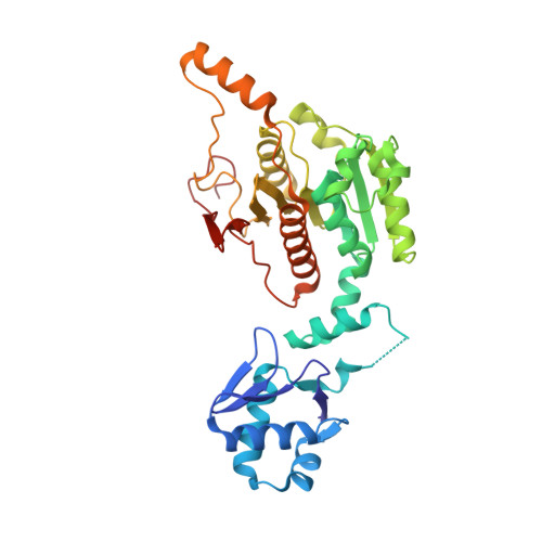
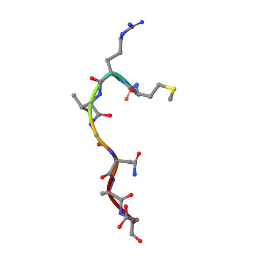
    6OM4

  • PubMed Abstract: 

    Microcin C7 (McC) is a peptide antibiotic modified by a linkage of the terminal isoAsn amide to AMP via a phosphoramidate bond. Post-translational modification on this ribosomally produced heptapeptide precursor is carried out by MccB, which consumes two equivalents of ATP to generate the N-P linkage. We demonstrate that MccB only efficiently processes the precursor heptapeptide that retains the N -formylated initiator Met (fMet). Binding studies and kinetic measurements evidence the role of the N -formyl moiety. Structural data show that the N -formyl peptide binding results in an ordering of residues in the MccB "crossover loop", which dictates specificity in homologous ubiquitin activating enzymes. The N -formyl peptide exhibits substrate inhibition, and cannot be displaced from MccB by the desformyl counterpart. Such substrate inhibition may be a strategy to avert unwanted McC buildup and avert toxicity in the cytoplasm of producing organisms.


  • Organizational Affiliation
    • Department of Biochemistry , University of Illinois at Urbana-Champaign , Illinois , USA . Email: snair@illinois.edu.

Macromolecule Content 

  • Total Structure Weight: 80.56 kDa 
  • Atom Count: 6,027 
  • Modeled Residue Count: 703 
  • Deposited Residue Count: 710 
  • Unique protein chains: 2

Macromolecules

Find similar proteins by:|  3D Structure
Entity ID: 1
MoleculeChains  Sequence LengthOrganismDetailsImage
MccB protein
A, B
348Escherichia coliMutation(s): 0 
Gene Names: mccB
UniProt
Find proteins for Q47506 (Escherichia coli)
Explore Q47506 
Go to UniProtKB:  Q47506
Entity Groups
Sequence Clusters30% Identity50% Identity70% Identity90% Identity95% Identity100% Identity
UniProt GroupQ47506
Sequence Annotations
Expand
Reference Sequence
Find similar proteins by:  Sequence   |   3D Structure  
Entity ID: 2
MoleculeChains  Sequence LengthOrganismDetailsImage
Microcin C7
C, D
7Escherichia coliMutation(s): 0 
UniProt
Find proteins for Q47505 (Escherichia coli)
Explore Q47505 
Go to UniProtKB:  Q47505
Entity Groups
UniProt GroupQ47505
Sequence Annotations
Expand
Reference Sequence

Small Molecules

Ligands 4 Unique
IDChains Name / Formula / InChI Key2D Diagram3D Interactions
ND7

Query on ND7


Download:Ideal Coordinates CCD File
M [auth C],
N [auth D]
5'-O-[(S)-amino(hydroxy)phosphoryl]adenosine
C10 H15 N6 O6 P
LDEMREUBLBGZBO-KQYNXXCUSA-N
POP

Query on POP


Download:Ideal Coordinates CCD File
G [auth A],
L [auth B]
PYROPHOSPHATE 2-
H2 O7 P2
XPPKVPWEQAFLFU-UHFFFAOYSA-L
ZN

Query on ZN


Download:Ideal Coordinates CCD File
E [auth A],
I [auth B]
ZINC ION
Zn
PTFCDOFLOPIGGS-UHFFFAOYSA-N
MG

Query on MG


Download:Ideal Coordinates CCD File
F [auth A],
H [auth A],
J [auth B],
K [auth B]
MAGNESIUM ION
Mg
JLVVSXFLKOJNIY-UHFFFAOYSA-N
Modified Residues  1 Unique
IDChains TypeFormula2D DiagramParent
FME
Query on FME
C, D
L-PEPTIDE LINKINGC6 H11 N O3 SMET

Experimental Data & Validation

Experimental Data

  • Method: X-RAY DIFFRACTION
  • Resolution: 1.70 Å
  • R-Value Free:  0.199 (Depositor), 0.206 (DCC) 
  • R-Value Work:  0.155 (Depositor), 0.165 (DCC) 
  • R-Value Observed: 0.157 (Depositor) 
Space Group: P 21 21 21
Unit Cell:
Length ( Å )Angle ( ˚ )
a = 55.094α = 90
b = 76.059β = 90
c = 131.902γ = 90
Software Package:
Software NamePurpose
REFMACrefinement
XDSdata scaling
PHENIXmodel building
BUCCANEERmodel building
PHENIXphasing
XDSdata reduction

Structure Validation

View Full Validation Report


Ligand Structure Quality Assessment 


Entry History 

& Funding Information

Deposition Data


Funding OrganizationLocationGrant Number
National Institutes of Health/National Human Genome Research Institute (NIH/NHGRI)United StatesAI117210

Revision History  (Full details and data files)

  • Version 1.0: 2019-05-22
    Type: Initial release
  • Version 1.1: 2019-12-18
    Changes: Author supporting evidence
  • Version 1.2: 2023-10-11
    Changes: Data collection, Database references, Derived calculations, Refinement description
  • Version 1.3: 2024-10-23
    Changes: Structure summary