6OJC | pdb_00006ojc

A high-resolution crystal structure of NocB thioesterase domain from Nocardicin cluster


Experimental Data Snapshot

  • Method: X-RAY DIFFRACTION
  • Resolution: 1.94 Å
  • R-Value Free: 
    0.230 (Depositor), 0.231 (DCC) 
  • R-Value Work: 
    0.206 (Depositor), 0.208 (DCC) 
  • R-Value Observed: 
    0.208 (Depositor) 

wwPDB Validation 3D Report Full Report

Validation slider image for 6OJC

This is version 1.2 of the entry. See complete history

Literature

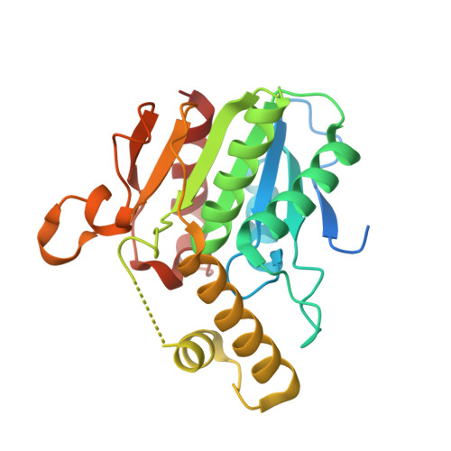
Structure of a bound peptide phosphonate reveals the mechanism of nocardicin bifunctional thioesterase epimerase-hydrolase half-reactions.

Patel, K.D.d'Andrea, F.B.Gaudelli, N.M.Buller, A.R.Townsend, C.A.Gulick, A.M.

(2019) Nat Commun 10: 3868-3868

  • DOI: https://doi.org/10.1038/s41467-019-11740-6
  • Primary Citation Related Structures: 
    6OJC, 6OJD

  • PubMed Abstract: 

    Nonribosomal peptide synthetases (NRPSs) underlie the biosynthesis of many natural products that have important medicinal utility. Protection of the NRPS peptide products from proteolysis is critical to these pathways and is often achieved by structural modification, principally the introduction of D-amino acid residues into the elongating peptide. These amino acids are generally formed in situ from their L-stereoisomers by epimerization domains or dual-function condensation/epimerization domains. In singular contrast, the thioesterase domain of nocardicin biosynthesis mediates both the effectively complete L- to D-epimerization of its C-terminal amino acid residue (≥100:1) and hydrolytic product release. We report herein high-resolution crystal structures of the nocardicin thioesterase domain in ligand-free form and reacted with a structurally precise fluorophosphonate substrate mimic that identify the complete peptide binding pocket to accommodate both stereoisomers. These structures combined with additional functional studies provide detailed mechanistic insight into this unique dual-function NRPS domain.


  • Organizational Affiliation
    • Department of Structural Biology, The Jacobs School of Medicine & Biomedical Sciences, State University of New York at Buffalo, 955 Main Street, Buffalo, NY, 14203, USA.

Macromolecule Content 

  • Total Structure Weight: 28.41 kDa 
  • Atom Count: 1,781 
  • Modeled Residue Count: 222 
  • Deposited Residue Count: 257 
  • Unique protein chains: 1

Macromolecules

Find similar proteins by:|  3D Structure
Entity ID: 1
MoleculeChains  Sequence LengthOrganismDetailsImage
NocB257Nocardia uniformis subsp. tsuyamanensisMutation(s): 0 
Gene Names: nocB
UniProt
Find proteins for Q5J1Q6 (Nocardia uniformis subsp. tsuyamanensis)
Explore Q5J1Q6 
Go to UniProtKB:  Q5J1Q6
Entity Groups
Sequence Clusters30% Identity50% Identity70% Identity90% Identity95% Identity100% Identity
UniProt GroupQ5J1Q6
Sequence Annotations
Expand
Reference Sequence

Experimental Data & Validation

Experimental Data

  • Method: X-RAY DIFFRACTION
  • Resolution: 1.94 Å
  • R-Value Free:  0.230 (Depositor), 0.231 (DCC) 
  • R-Value Work:  0.206 (Depositor), 0.208 (DCC) 
  • R-Value Observed: 0.208 (Depositor) 
Space Group: P 3 2 1
Unit Cell:
Length ( Å )Angle ( ˚ )
a = 114.73α = 90
b = 114.73β = 90
c = 46.601γ = 120
Software Package:
Software NamePurpose
PHENIXrefinement
XDSdata reduction
Aimlessdata scaling
PHASERphasing

Structure Validation

View Full Validation Report


Entry History 

& Funding Information

Deposition Data


Funding OrganizationLocationGrant Number
National Institutes of Health/National Human Genome Research Institute (NIH/NHGRI)United StatesR01GM116957

Revision History  (Full details and data files)

  • Version 1.0: 2019-09-11
    Type: Initial release
  • Version 1.1: 2019-12-18
    Changes: Author supporting evidence
  • Version 1.2: 2024-03-13
    Changes: Advisory, Data collection, Database references