6OB6 | pdb_00006ob6

Human equilibrative nucleoside transporter-1, S-(4-nitrobenzyl)-6-thioinosine bound, merohedrally twinned


Experimental Data Snapshot

  • Method: X-RAY DIFFRACTION
  • Resolution: 2.90 Å
  • R-Value Free: 
    0.252 (Depositor), 0.248 (DCC) 
  • R-Value Work: 
    0.205 (Depositor), 0.199 (DCC) 
  • R-Value Observed: 
    0.214 (Depositor) 

Starting Model: experimental
View more details

wwPDB Validation 3D Report Full Report

Validation slider image for 6OB6

Ligand Structure Quality Assessment 


This is version 2.2 of the entry. See complete history

Literature

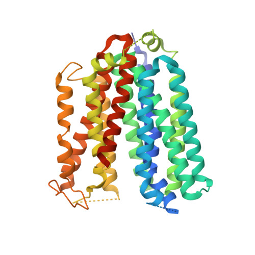
Structures of human ENT1 in complex with adenosine reuptake inhibitors.

Wright, N.J.Lee, S.Y.

(2019) Nat Struct Mol Biol 26: 599-606

  • DOI: https://doi.org/10.1038/s41594-019-0245-7
  • Primary Citation Related Structures: 
    6OB6, 6OB7

  • PubMed Abstract: 

    The human equilibrative nucleoside transporter 1 (hENT1), a member of the SLC29 family, plays crucial roles in adenosine signaling, cellular uptake of nucleoside for DNA and RNA synthesis, and nucleoside-derived anticancer and antiviral drug transport in humans. Because of its central role in adenosine signaling, it is the target of adenosine reuptake inhibitors (AdoRI), several of which are used clinically. Despite its importance in human physiology and pharmacology, the molecular basis of hENT1-mediated adenosine transport and its inhibition by AdoRIs are limited, owing to the absence of structural information on hENT1. Here, we present crystal structures of hENT1 in complex with two chemically distinct AdoRIs: dilazep and S-(4-nitrobenzyl)-6-thioinosine (NBMPR). Combined with mutagenesis study, our structural analyses elucidate two distinct inhibitory mechanisms exhibited on hENT1 and provide insight into adenosine recognition and transport. Our studies provide a platform for improved pharmacological intervention of adenosine and nucleoside analog drug transport by hENT1.


  • Organizational Affiliation
    • Department of Biochemistry, Duke University Medical Center, Durham, NC, USA.

Macromolecule Content 

  • Total Structure Weight: 98.57 kDa 
  • Atom Count: 5,512 
  • Modeled Residue Count: 722 
  • Deposited Residue Count: 884 
  • Unique protein chains: 1

Macromolecules

Find similar proteins by:|  3D Structure
Entity ID: 1
MoleculeChains  Sequence LengthOrganismDetailsImage
Equilibrative nucleoside transporter 1
A, B
442Homo sapiensMutation(s): 3 
Gene Names: SLC29A1ENT1
Membrane Entity: Yes 
UniProt & NIH Common Fund Data Resources
Find proteins for Q99808 (Homo sapiens)
Explore Q99808 
Go to UniProtKB:  Q99808
PHAROS:  Q99808
GTEx:  ENSG00000112759 
Entity Groups
Sequence Clusters30% Identity50% Identity70% Identity90% Identity95% Identity100% Identity
UniProt GroupQ99808
Sequence Annotations
Expand
Reference Sequence

Small Molecules

Ligands 1 Unique
IDChains Name / Formula / InChI Key2D Diagram3D Interactions
NBM

Query on NBM


Download:Ideal Coordinates CCD File
C [auth A],
D [auth B]
6-{[(4-nitrophenyl)methyl]sulfanyl}-9-beta-D-ribofuranosyl-9H-purine
C17 H17 N5 O6 S
DYCJFJRCWPVDHY-LSCFUAHRSA-N
Binding Affinity Annotations 
IDSourceBinding Affinity
NBM BindingDB:  6OB6 Ki: min: 0.43, max: 0.71 (nM) from 3 assay(s)
IC50: min: 1, max: 8.51 (nM) from 3 assay(s)

Experimental Data & Validation

Experimental Data

  • Method: X-RAY DIFFRACTION
  • Resolution: 2.90 Å
  • R-Value Free:  0.252 (Depositor), 0.248 (DCC) 
  • R-Value Work:  0.205 (Depositor), 0.199 (DCC) 
  • R-Value Observed: 0.214 (Depositor) 
Space Group: P 61
Unit Cell:
Length ( Å )Angle ( ˚ )
a = 72.532α = 90
b = 72.532β = 90
c = 335.458γ = 120
Software Package:
Software NamePurpose
PHENIXrefinement
XDSdata reduction
Aimlessdata scaling
STARANISOdata scaling
PHENIXphasing

Structure Validation

View Full Validation Report


Ligand Structure Quality Assessment 


Entry History 

& Funding Information

Deposition Data


Funding OrganizationLocationGrant Number
National Institutes of Health/National Institute of Neurological Disorders and Stroke (NIH/NINDS)United StatesR35NS097241

Revision History  (Full details and data files)

  • Version 1.0: 2019-07-03
    Type: Initial release
  • Version 1.1: 2019-07-10
    Changes: Data collection, Database references
  • Version 1.2: 2019-07-17
    Changes: Data collection, Database references
  • Version 2.0: 2019-09-18
    Changes: Advisory, Atomic model, Data collection, Database references, Derived calculations, Polymer sequence, Refinement description, Source and taxonomy, Structure summary
  • Version 2.1: 2019-12-18
    Changes: Author supporting evidence
  • Version 2.2: 2023-10-11
    Changes: Data collection, Database references, Refinement description