6O65 | pdb_00006o65

Crystal Structure of Arabidopsis thaliana Spermidine Synthase isoform 1 (AtSPDS1) in complex with decarboxylated S-adenosylmethionine and cyclohexylamine


Experimental Data Snapshot

  • Method: X-RAY DIFFRACTION
  • Resolution: 1.80 Å
  • R-Value Free: 
    0.261 (Depositor), 0.265 (DCC) 
  • R-Value Work: 
    0.219 (Depositor), 0.225 (DCC) 
  • R-Value Observed: 
    0.219 (Depositor) 

wwPDB Validation 3D Report Full Report

Validation slider image for 6O65

Ligand Structure Quality Assessment 


This is version 1.2 of the entry. See complete history

Literature

Spermidine Synthase (SPDS) Undergoes Concerted Structural Rearrangements Upon Ligand Binding - A Case Study of the Two SPDS Isoforms FromArabidopsis thaliana.

Sekula, B.Dauter, Z.

(2019) Front Plant Sci 10: 555-555

  • DOI: https://doi.org/10.3389/fpls.2019.00555
  • Primary Citation Related Structures: 
    6O63, 6O64, 6O65

  • PubMed Abstract: 

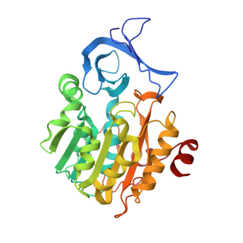
    Spermidine synthases (SPDSs) catalyze the production of the linear triamine, spermidine, from putrescine. They utilize decarboxylated S-adenosylmethionine (dc-SAM), a universal cofactor of aminopropyltransferases, as a donor of the aminopropyl moiety. In this work, we describe crystal structures of two SPDS isoforms from Arabidopsis thaliana ( At SPDS1 and At SPDS2). At SPDS1 and At SPDS2 are dimeric enzymes that share the fold of the polyamine biosynthesis proteins. Subunits of both isoforms present the characteristic two-domain structure. Smaller, N-terminal domain is built of the two β-sheets, while the C-terminal domain has a Rossmann fold-like topology. The catalytic cleft composed of two main compartments, the dc-SAM binding site and the polyamine groove, is created independently in each At SPDS subunits at the domain interface. We also provide the structural details about the dc-SAM binding mode and the inhibition of SPDS by a potent competitive inhibitor, cyclohexylamine (CHA). CHA occupies the polyamine binding site of At SPDS where it is bound at the bottom of the active site with the amine group placed analogously to the substrate. The crystallographic snapshots show in detail the structural rearrangements of At SPDS1 and At SPDS2 that are required to stabilize ligands within the active site. The concerted movements are observed in both compartments of the catalytic cleft, where three major parts significantly change their conformation. These are (i) the neighborhood of the glycine-rich region where aminopropyl moiety of dc-SAM is bound, (ii) the very flexible gate region with helix η6, which interacts with both, the adenine moiety of dc-SAM and the bound polyamine or inhibitor, and (iii) the N-terminal β-hairpin, that limits the putrescine binding grove at the bottom of the catalytic site.


  • Organizational Affiliation
    • Synchrotron Radiation Research Section, Macromolecular Crystallography Laboratory, National Cancer Institute, Argonne, IL, United States.

Macromolecule Content 

  • Total Structure Weight: 270.72 kDa 
  • Atom Count: 20,468 
  • Modeled Residue Count: 2,333 
  • Deposited Residue Count: 2,432 
  • Unique protein chains: 1

Macromolecules

Find similar proteins by:|  3D Structure
Entity ID: 1
MoleculeChains  Sequence LengthOrganismDetailsImage
Spermidine synthase 1
A, B, C, D, E
A, B, C, D, E, F, G, H
304Arabidopsis thalianaMutation(s): 0 
Gene Names: SPDSYN1At1g23820F5O8.38
EC: 2.5.1.16
UniProt
Find proteins for Q9ZUB3 (Arabidopsis thaliana)
Explore Q9ZUB3 
Go to UniProtKB:  Q9ZUB3
Entity Groups
Sequence Clusters30% Identity50% Identity70% Identity90% Identity95% Identity100% Identity
UniProt GroupQ9ZUB3
Sequence Annotations
Expand
Reference Sequence

Small Molecules

Ligands 5 Unique
IDChains Name / Formula / InChI Key2D Diagram3D Interactions
S4M

Query on S4M


Download:Ideal Coordinates CCD File
EA [auth H]
I [auth A]
K [auth B]
N [auth C]
Q [auth D]
EA [auth H],
I [auth A],
K [auth B],
N [auth C],
Q [auth D],
S [auth E],
W [auth F],
Z [auth G]
5'-[(S)-(3-AMINOPROPYL)(METHYL)-LAMBDA~4~-SULFANYL]-5'-DEOXYADENOSINE
C14 H24 N6 O3 S
KIZZALRCMBIHBH-IDTAVKCVSA-N
PEG

Query on PEG


Download:Ideal Coordinates CCD File
BA [auth G],
U [auth E]
DI(HYDROXYETHYL)ETHER
C4 H10 O3
MTHSVFCYNBDYFN-UHFFFAOYSA-N
HAI

Query on HAI


Download:Ideal Coordinates CCD File
AA [auth G]
FA [auth H]
J [auth A]
L [auth B]
O [auth C]
AA [auth G],
FA [auth H],
J [auth A],
L [auth B],
O [auth C],
R [auth D],
T [auth E],
X [auth F]
CYCLOHEXYLAMMONIUM ION
C6 H14 N
PAFZNILMFXTMIY-UHFFFAOYSA-O
SO4

Query on SO4


Download:Ideal Coordinates CCD File
DA [auth G],
M [auth B],
P [auth C],
V [auth E]
SULFATE ION
O4 S
QAOWNCQODCNURD-UHFFFAOYSA-L
EDO

Query on EDO


Download:Ideal Coordinates CCD File
CA [auth G],
Y [auth F]
1,2-ETHANEDIOL
C2 H6 O2
LYCAIKOWRPUZTN-UHFFFAOYSA-N

Experimental Data & Validation

Experimental Data

  • Method: X-RAY DIFFRACTION
  • Resolution: 1.80 Å
  • R-Value Free:  0.261 (Depositor), 0.265 (DCC) 
  • R-Value Work:  0.219 (Depositor), 0.225 (DCC) 
  • R-Value Observed: 0.219 (Depositor) 
Space Group: P 1 21 1
Unit Cell:
Length ( Å )Angle ( ˚ )
a = 89.201α = 90
b = 107.558β = 95.3
c = 142.424γ = 90
Software Package:
Software NamePurpose
REFMACrefinement
PHASERphasing
STARANISOdata scaling
XDSdata reduction

Structure Validation

View Full Validation Report


Ligand Structure Quality Assessment 


Entry History 

& Funding Information

Deposition Data


Funding OrganizationLocationGrant Number
National Institutes of Health/National Cancer Institute (NIH/NCI)United StatesThe Intramural Research Program

Revision History  (Full details and data files)

  • Version 1.0: 2019-06-05
    Type: Initial release
  • Version 1.1: 2019-12-04
    Changes: Author supporting evidence
  • Version 1.2: 2024-03-13
    Changes: Data collection, Database references