Parsing the functional specificity of Siderocalin/Lipocalin 2/NGAL for siderophores and related small-molecule ligands.
Clifton, M.C., Rupert, P.B., Hoette, T.M., Raymond, K.N., Abergel, R.J., Strong, R.K.(2019) J Struct Biol X 2: 100008-100008
- PubMed: 32647813 Search on PubMedSearch on PubMed Central
- DOI: https://doi.org/10.1016/j.yjsbx.2019.100008
- Primary Citation Related Structures:
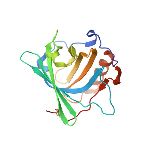
3T1D, 6O5D - PubMed Abstract:
Siderocalin/Lipocalin 2/Neutrophil Gelatinase Associated Lipocalin/24p3 is an innate immune system protein with bacteriostatic activity, acting by tightly binding and sequestering diverse catecholate and mixed-type ferric siderophores from enteric bacteria and mycobacteria. Bacterial virulence achieved through siderophore modifications, or utilization of alternate siderophores, can be explained by evasion of Siderocalin binding. Siderocalin has also been implicated in a wide variety of disease processes, though often in seemingly contradictory ways, and has been proposed to bind to a broader array of ligands beyond siderophores. Using structural, directed mutational, and binding studies, we have sought to rigorously test, and fully elucidate, the Siderocalin recognition mechanism. Several proposed ligands fail to meet rigorous binding criteria, including the bacterial siderophore pyochelin, the iron-chelating catecholamine hormone norepinephrine, and the bacterial second messenger cyclic diguanylate monophosphate. While possessing a remarkably rigid structure, in principle simplifying analyses of ligand recognition, understanding Scn recognition is complicated by the observed conformational and stoichiometric plasticity, and instability, of its bona fide siderophore ligands. Since the role of Siderocalin at the early host/pathogen interface is to compete for bacterial ferric siderophores, we also analyzed how bacterial siderophore binding proteins and enzymes alternately recognize siderophores that efficiently bind to, or evade, Siderocalin sequestration - including determining the crystal structure of Bacillus cereus YfiY bound to schizokinen. These studies combine to refine the potential physiological functions of Siderocalin by defining its multiplexed recognition mechanism.
- Division of Basic Science, Fred Hutchinson Cancer Research Center, Seattle, WA 98109, USA.
Organizational Affiliation: