6O55 | pdb_00006o55

Crystal Structure of N5-carboxyaminoimidazole ribonucleotide mutase (PurE) from Legionella pneumophila


Experimental Data Snapshot

  • Method: X-RAY DIFFRACTION
  • Resolution: 1.70 Å
  • R-Value Free: 
    0.182 (Depositor), 0.181 (DCC) 
  • R-Value Work: 
    0.148 (Depositor), 0.147 (DCC) 
  • R-Value Observed: 
    0.149 (Depositor) 

Starting Model: experimental
View more details

wwPDB Validation 3D Report Full Report

Validation slider image for 6O55

This is version 1.3 of the entry. See complete history

Literature

Structure-Guided Discovery of N 5 -CAIR Mutase Inhibitors.

Belfon, K.K.J.Sharma, N.Zigweid, R.Bolejack, M.Davies, D.Edwards, T.E.Myler, P.J.French, J.B.

(2023) Biochemistry 62: 2587-2596

  • DOI: https://doi.org/10.1021/acs.biochem.2c00705
  • Primary Citation Related Structures: 
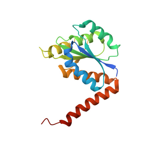
    4GRD, 6O55

  • PubMed Abstract: 

    Because purine nucleotides are essential for all life, differences between how microbes and humans metabolize purines can be exploited for the development of antimicrobial therapies. While humans biosynthesize purine nucleotides in a 10-step pathway, most microbes utilize an additional 11th enzymatic activity. The human enzyme, aminoimidazole ribonucleotide (AIR) carboxylase generates the product 4-carboxy-5-aminoimidazole ribonucleotide (CAIR) directly. Most microbes, however, require two separate enzymes, a synthetase (PurK) and a mutase (PurE), and proceed through the intermediate, N 5 -CAIR. Toward the development of therapeutics that target these differences, we have solved crystal structures of the N 5 -CAIR mutase of the human pathogens Legionella pneumophila (LpPurE) and Burkholderia cenocepacia (BcPurE) and used a structure-guided approach to identify inhibitors. Analysis of the structures reveals a highly conserved fold and active site architecture. Using this data, and three additional structures of PurE enzymes, we screened a library of FDA-approved compounds in silico and identified a set of 25 candidates for further analysis. Among these, we identified several new PurE inhibitors with micromolar IC 50 values. Several of these compounds, including the α 1 -blocker Alfuzosin, inhibit the microbial PurE enzymes much more effectively than the human homologue. These structures and the newly described PurE inhibitors are valuable tools to aid in further studies of this enzyme and provide a foundation for the development of compounds that target differences between human and microbial purine metabolism.


  • Organizational Affiliation
    • Department of Biochemistry and Cell Biology, Stony Brook University, Stony Brook, New York 11794, United States.

Macromolecule Content 

  • Total Structure Weight: 75.02 kDa 
  • Atom Count: 5,519 
  • Modeled Residue Count: 646 
  • Deposited Residue Count: 696 
  • Unique protein chains: 1

Macromolecules

Find similar proteins by:|  3D Structure
Entity ID: 1
MoleculeChains  Sequence LengthOrganismDetailsImage
N5-carboxyaminoimidazole ribonucleotide mutase
A, B, C, D
174Legionella pneumophila subsp. pneumophila str. Philadelphia 1Mutation(s): 0 
Gene Names: purElpg0218
EC: 5.4.99.18
UniProt
Find proteins for Q5ZYZ3 (Legionella pneumophila subsp. pneumophila (strain Philadelphia 1 / ATCC 33152 / DSM 7513))
Explore Q5ZYZ3 
Go to UniProtKB:  Q5ZYZ3
Entity Groups
Sequence Clusters30% Identity50% Identity70% Identity90% Identity95% Identity100% Identity
UniProt GroupQ5ZYZ3
Sequence Annotations
Expand
Reference Sequence

Small Molecules

Ligands 3 Unique
IDChains Name / Formula / InChI Key2D Diagram3D Interactions
PG4

Query on PG4


Download:Ideal Coordinates CCD File
K [auth C]TETRAETHYLENE GLYCOL
C8 H18 O5
UWHCKJMYHZGTIT-UHFFFAOYSA-N
EDO

Query on EDO


Download:Ideal Coordinates CCD File
E [auth A]
F [auth A]
H [auth B]
I [auth B]
M [auth D]
E [auth A],
F [auth A],
H [auth B],
I [auth B],
M [auth D],
N [auth D]
1,2-ETHANEDIOL
C2 H6 O2
LYCAIKOWRPUZTN-UHFFFAOYSA-N
CL

Query on CL


Download:Ideal Coordinates CCD File
G [auth A],
J [auth B],
L [auth C],
O [auth D]
CHLORIDE ION
Cl
VEXZGXHMUGYJMC-UHFFFAOYSA-M

Experimental Data & Validation

Experimental Data

  • Method: X-RAY DIFFRACTION
  • Resolution: 1.70 Å
  • R-Value Free:  0.182 (Depositor), 0.181 (DCC) 
  • R-Value Work:  0.148 (Depositor), 0.147 (DCC) 
  • R-Value Observed: 0.149 (Depositor) 
Space Group: C 1 2 1
Unit Cell:
Length ( Å )Angle ( ˚ )
a = 137.28α = 90
b = 88.56β = 111.77
c = 52.51γ = 90
Software Package:
Software NamePurpose
PHENIXrefinement
XDSdata reduction
XSCALEdata scaling
PDB_EXTRACTdata extraction
MoRDaphasing

Structure Validation

View Full Validation Report


Entry History 

Deposition Data

Revision History  (Full details and data files)

  • Version 1.0: 2019-03-13
    Type: Initial release
  • Version 1.1: 2023-10-11
    Changes: Data collection, Database references, Refinement description
  • Version 1.2: 2024-05-29
    Changes: Database references
  • Version 1.3: 2025-10-22
    Changes: Structure summary