6O3A | pdb_00006o3a

Crystal structure of Frizzled 7 CRD in complex with F7.B Fab


Experimental Data Snapshot

  • Method: X-RAY DIFFRACTION
  • Resolution: 2.10 Å
  • R-Value Free: 
    0.242 (Depositor), 0.239 (DCC) 
  • R-Value Work: 
    0.192 (Depositor), 0.194 (DCC) 
  • R-Value Observed: 
    0.194 (Depositor) 

Starting Model: experimental
View more details

wwPDB Validation 3D Report Full Report

Validation slider image for 6O3A

Ligand Structure Quality Assessment 


This is version 1.4 of the entry. See complete history

Literature

Structure-guided design fine-tunes pharmacokinetics, tolerability, and antitumor profile of multispecific frizzled antibodies.

Raman, S.Beilschmidt, M.To, M.Lin, K.Lui, F.Jmeian, Y.Ng, M.Fernandez, M.Fu, Y.Mascall, K.Duque, A.Wang, X.Pan, G.Angers, S.Moffat, J.Sidhu, S.S.Magram, J.Sinclair, A.M.Fransson, J.Julien, J.P.

(2019) Proc Natl Acad Sci U S A 116: 6812-6817

  • DOI: https://doi.org/10.1073/pnas.1817246116
  • Primary Citation Related Structures: 
    6O39, 6O3A, 6O3B

  • PubMed Abstract: 

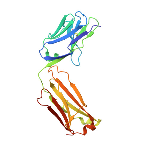
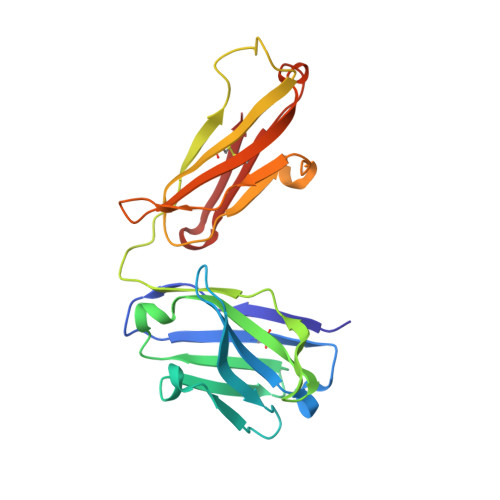
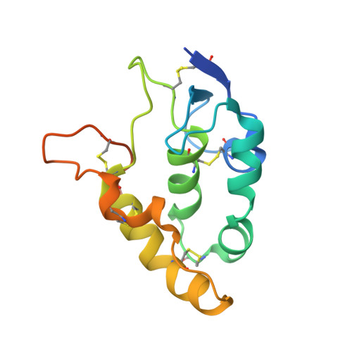
    Aberrant activation of Wnt/β-catenin signaling occurs frequently in cancer. However, therapeutic targeting of this pathway is complicated by the role of Wnt in stem cell maintenance and tissue homeostasis. Here, we evaluated antibodies blocking 6 of the 10 human Wnt/Frizzled (FZD) receptors as potential therapeutics. Crystal structures revealed a common binding site for these monoclonal antibodies (mAbs) on FZD, blocking the interaction with the Wnt palmitoleic acid moiety. However, these mAbs displayed gastrointestinal toxicity or poor plasma exposure in vivo. Structure-guided engineering was used to refine the binding of each mAb for FZD receptors, resulting in antibody variants with improved in vivo tolerability and developability. Importantly, the lead variant mAb significantly inhibited tumor growth in the HPAF-II pancreatic tumor xenograft model. Taken together, our data demonstrate that anti-FZD cancer therapeutic antibodies with broad specificity can be fine-tuned to navigate in vivo exposure and tolerability while driving therapeutic efficacy.


  • Organizational Affiliation
    • Program in Molecular Medicine, Hospital for Sick Children Research Institute, Toronto, ON M5G 0A4, Canada.

Macromolecule Content 

  • Total Structure Weight: 62.73 kDa 
  • Atom Count: 4,583 
  • Modeled Residue Count: 548 
  • Deposited Residue Count: 568 
  • Unique protein chains: 3

Macromolecules

Find similar proteins by:|  3D Structure
Entity ID: 1
MoleculeChains  Sequence LengthOrganismDetailsImage
Antibody F7.B Fab, Light chain212Homo sapiensMutation(s): 0 
Find similar proteins by:|  3D Structure
Entity ID: 2
MoleculeChains  Sequence LengthOrganismDetailsImage
Antibody F7.B Fab, Heavy chain218Homo sapiensMutation(s): 0 
Find similar proteins by:|  3D Structure
Entity ID: 3
MoleculeChains  Sequence LengthOrganismDetailsImage
Frizzled-7C [auth E]138Homo sapiensMutation(s): 0 
Gene Names: FZD7
UniProt & NIH Common Fund Data Resources
Find proteins for O75084 (Homo sapiens)
Explore O75084 
Go to UniProtKB:  O75084
PHAROS:  O75084
GTEx:  ENSG00000155760 
Entity Groups
Sequence Clusters30% Identity50% Identity70% Identity90% Identity95% Identity100% Identity
UniProt GroupO75084
Glycosylation
Glycosylation Sites: 1Go to GlyGen: O75084-1
Sequence Annotations
Expand
Reference Sequence

Small Molecules

Ligands 4 Unique
IDChains Name / Formula / InChI Key2D Diagram3D Interactions
CXS

Query on CXS


Download:Ideal Coordinates CCD File
D [auth A]3-CYCLOHEXYL-1-PROPYLSULFONIC ACID
C9 H19 N O3 S
PJWWRFATQTVXHA-UHFFFAOYSA-N
NAG

Query on NAG


Download:Ideal Coordinates CCD File
J [auth B],
M [auth E]
2-acetamido-2-deoxy-beta-D-glucopyranose
C8 H15 N O6
OVRNDRQMDRJTHS-FMDGEEDCSA-N
EDO

Query on EDO


Download:Ideal Coordinates CCD File
E [auth A]
F [auth A]
G [auth A]
H [auth A]
K [auth B]
E [auth A],
F [auth A],
G [auth A],
H [auth A],
K [auth B],
L [auth B]
1,2-ETHANEDIOL
C2 H6 O2
LYCAIKOWRPUZTN-UHFFFAOYSA-N
NA

Query on NA


Download:Ideal Coordinates CCD File
I [auth A]SODIUM ION
Na
FKNQFGJONOIPTF-UHFFFAOYSA-N

Experimental Data & Validation

Experimental Data

  • Method: X-RAY DIFFRACTION
  • Resolution: 2.10 Å
  • R-Value Free:  0.242 (Depositor), 0.239 (DCC) 
  • R-Value Work:  0.192 (Depositor), 0.194 (DCC) 
  • R-Value Observed: 0.194 (Depositor) 
Space Group: P 21 21 21
Unit Cell:
Length ( Å )Angle ( ˚ )
a = 41.544α = 90
b = 103.446β = 90
c = 144.207γ = 90
Software Package:
Software NamePurpose
PHENIXrefinement
XPREPdata reduction
XDSdata scaling
PHENIXphasing

Structure Validation

View Full Validation Report


Ligand Structure Quality Assessment 


Entry History 

Deposition Data

Revision History  (Full details and data files)

  • Version 1.0: 2019-04-03
    Type: Initial release
  • Version 1.1: 2019-04-17
    Changes: Data collection, Database references
  • Version 1.2: 2020-07-29
    Type: Remediation
    Reason: Carbohydrate remediation
    Changes: Data collection, Derived calculations, Structure summary
  • Version 1.3: 2023-10-11
    Changes: Data collection, Database references, Refinement description, Structure summary
  • Version 1.4: 2024-11-20
    Changes: Structure summary