6O0H | pdb_00006o0h

Cryo-EM structure of human ATP-citrate lyase in complex with inhibitor NDI-091143

  • Classification: ligase/ligase inhibitor
  • Organism(s): Homo sapiens
  • Expression System: Escherichia coli
  • Mutation(s): No 

  • Deposited: 2019-02-16 Released: 2019-04-17 
  • Deposition Author(s): Wei, J., Tong, L.
  • Funding Organization(s): Other private, National Institutes of Health/National Institute of General Medical Sciences (NIH/NIGMS), Other government

Experimental Data Snapshot

  • Method: ELECTRON MICROSCOPY
  • Resolution: 3.67 Å
  • Aggregation State: PARTICLE 
  • Reconstruction Method: SINGLE PARTICLE 

wwPDB Validation 3D Report Full Report

Validation slider image for 6O0H

This is version 1.3 of the entry. See complete history

Literature

An allosteric mechanism for potent inhibition of human ATP-citrate lyase.

Wei, J.Leit, S.Kuai, J.Therrien, E.Rafi, S.Harwood Jr., H.J.DeLaBarre, B.Tong, L.

(2019) Nature 568: 566-570

  • DOI: https://doi.org/10.1038/s41586-019-1094-6
  • Primary Citation Related Structures: 
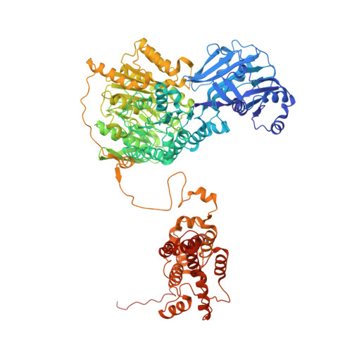
    6O0H

  • PubMed Abstract: 

    ATP-citrate lyase (ACLY) is a central metabolic enzyme and catalyses the ATP-dependent conversion of citrate and coenzyme A (CoA) to oxaloacetate and acetyl-CoA 1-5 . The acetyl-CoA product is crucial for the metabolism of fatty acids 6,7 , the biosynthesis of cholesterol 8 , and the acetylation and prenylation of proteins 9,10 . There has been considerable interest in ACLY as a target for anti-cancer drugs, because many cancer cells depend on its activity for proliferation 2,5,11 . ACLY is also a target against dyslipidaemia and hepatic steatosis, with a compound currently in phase 3 clinical trials 4,5 . Many inhibitors of ACLY have been reported, but most of them have weak activity 5 . Here we report the development of a series of low nanomolar, small-molecule inhibitors of human ACLY. We have also determined the structure of the full-length human ACLY homo-tetramer in complex with one of these inhibitors (NDI-091143) by cryo-electron microscopy, which reveals an unexpected mechanism of inhibition. The compound is located in an allosteric, mostly hydrophobic cavity next to the citrate-binding site, and requires extensive conformational changes in the enzyme that indirectly disrupt citrate binding. The observed binding mode is supported by and explains the structure-activity relationships of these compounds. This allosteric site greatly enhances the 'druggability' of ACLY and represents an attractive target for the development of new ACLY inhibitors.


  • Organizational Affiliation
    • Department of Biological Sciences, Columbia University, New York, NY, USA.

Macromolecule Content 

  • Total Structure Weight: 494.67 kDa 
  • Atom Count: 32,012 
  • Modeled Residue Count: 4,092 
  • Deposited Residue Count: 4,464 
  • Unique protein chains: 1

Macromolecules

Find similar proteins by:|  3D Structure
Entity ID: 1
MoleculeChains  Sequence LengthOrganismDetailsImage
ATP-citrate synthase
A, B, C, D
1,116Homo sapiensMutation(s): 0 
Gene Names: ACLY
EC: 2.3.3.8
UniProt & NIH Common Fund Data Resources
Find proteins for P53396 (Homo sapiens)
Explore P53396 
Go to UniProtKB:  P53396
PHAROS:  P53396
GTEx:  ENSG00000131473 
Entity Groups
Sequence Clusters30% Identity50% Identity70% Identity90% Identity95% Identity100% Identity
UniProt GroupP53396
Sequence Annotations
Expand
Reference Sequence

Small Molecules

Ligands 2 Unique
IDChains Name / Formula / InChI Key2D Diagram3D Interactions
LBG

Query on LBG


Download:Ideal Coordinates CCD File
E [auth A],
G [auth B],
I [auth C],
K [auth D]
methyl 3-chloro-5-[(4,6-difluoro[1,1'-biphenyl]-3-yl)sulfamoyl]-4-hydroxybenzoate
C20 H14 Cl F2 N O5 S
YSTSHUWHIDBZAK-UHFFFAOYSA-N
ADP

Query on ADP


Download:Ideal Coordinates CCD File
F [auth A],
H [auth B],
J [auth C],
L [auth D]
ADENOSINE-5'-DIPHOSPHATE
C10 H15 N5 O10 P2
XTWYTFMLZFPYCI-KQYNXXCUSA-N

Experimental Data & Validation

Experimental Data

  • Method: ELECTRON MICROSCOPY
  • Resolution: 3.67 Å
  • Aggregation State: PARTICLE 
  • Reconstruction Method: SINGLE PARTICLE 
EM Software:
TaskSoftware PackageVersion
RECONSTRUCTIONRELION3.0-beta-2
MODEL REFINEMENTPHENIX

Structure Validation

View Full Validation Report


Entry History 

& Funding Information

Deposition Data


Funding OrganizationLocationGrant Number
Other privateUnited StatesNimbus Therapeutics
Other privateUnited StatesSimons Foundation (349247)
National Institutes of Health/National Institute of General Medical Sciences (NIH/NIGMS)United StatesGM103310
Other governmentUnited StatesNYSTAR

Revision History  (Full details and data files)

  • Version 1.0: 2019-04-17
    Type: Initial release
  • Version 1.1: 2019-05-08
    Changes: Data collection, Database references
  • Version 1.2: 2020-01-08
    Changes: Author supporting evidence, Data collection
  • Version 1.3: 2024-03-20
    Changes: Data collection, Database references