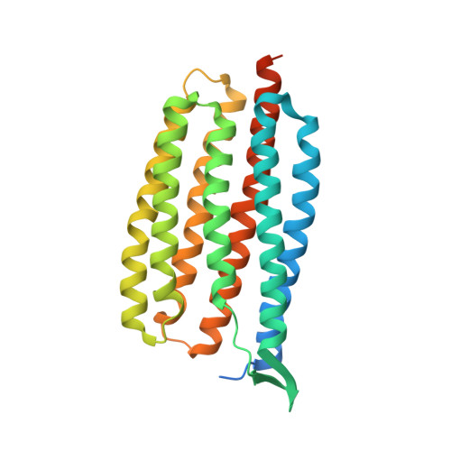
6NWD

X-ray Crystallographic structure of Gloeobacter rhodopsin


Experimental Data Snapshot

  • Method: X-RAY DIFFRACTION
  • Resolution: 2.00 Å
  • R-Value Free: 0.244 
  • R-Value Work: 0.230 

Starting Model: experimental
View more details

wwPDB Validation   3D Report Full Report


Ligand Structure Quality Assessment 


This is version 1.2 of the entry. See complete history


Literature

X-ray Crystallographic Structure and Oligomerization of Gloeobacter Rhodopsin.

Morizumi, T.Ou, W.L.Van Eps, N.Inoue, K.Kandori, H.Brown, L.S.Ernst, O.P.

(2019) Sci Rep 9: 11283-11283

  • DOI: https://doi.org/10.1038/s41598-019-47445-5
  • Primary Citation of Related Structures:  
    6NWD

  • PubMed Abstract: 

    Gloeobacter rhodopsin (GR) is a cyanobacterial proton pump which can be potentially applied to optogenetics. We solved the crystal structure of GR and found that it has overall similarity to the homologous proton pump from Salinibacter ruber, xanthorhodopsin (XR). We identified distinct structural characteristics of GR's hydrogen bonding network in the transmembrane domain as well as the displacement of extracellular sides of the transmembrane helices relative to those of XR. Employing Raman spectroscopy and flash-photolysis, we found that GR in the crystals exists in a state which displays retinal conformation and photochemical cycle similar to the functional form observed in lipids. Based on the crystal structure of GR, we selected a site for spin labeling to determine GR's oligomerization state using double electron-electron resonance (DEER) spectroscopy and demonstrated the pH-dependent pentamer formation of GR. Determination of the structure of GR as well as its pentamerizing propensity enabled us to reveal the role of structural motifs (extended helices, 3-omega motif and flipped B-C loop) commonly found among light-driven bacterial pumps in oligomer formation. Here we propose a new concept to classify these pumps based on the relationship between their oligomerization propensities and these structural determinants.


  • Organizational Affiliation

    Department of Biochemistry, University of Toronto, Toronto, Ontario, M5S 1A8, Canada.


Macromolecules
Find similar proteins by:  (by identity cutoff)  |  3D Structure
Entity ID: 1
MoleculeChains Sequence LengthOrganismDetailsImage
Gll0198 protein298Gloeobacter violaceus PCC 7421Mutation(s): 0 
Gene Names: gll0198
Membrane Entity: Yes 
UniProt
Find proteins for Q7NP59 (Gloeobacter violaceus (strain ATCC 29082 / PCC 7421))
Explore Q7NP59 
Go to UniProtKB:  Q7NP59
Entity Groups  
Sequence Clusters30% Identity50% Identity70% Identity90% Identity95% Identity100% Identity
UniProt GroupQ7NP59
Sequence Annotations
Expand
  • Reference Sequence
Small Molecules
Ligands 4 Unique
IDChains Name / Formula / InChI Key2D Diagram3D Interactions
PX4
Query on PX4

Download Ideal Coordinates CCD File 
C [auth A],
D [auth A],
E [auth A]
1,2-DIMYRISTOYL-SN-GLYCERO-3-PHOSPHOCHOLINE
C36 H73 N O8 P
CITHEXJVPOWHKC-UUWRZZSWSA-O
RET (Subject of Investigation/LOI)
Query on RET

Download Ideal Coordinates CCD File 
B [auth A]RETINAL
C20 H28 O
NCYCYZXNIZJOKI-OVSJKPMPSA-N
D12
Query on D12

Download Ideal Coordinates CCD File 
G [auth A]DODECANE
C12 H26
SNRUBQQJIBEYMU-UHFFFAOYSA-N
D10
Query on D10

Download Ideal Coordinates CCD File 
F [auth A]DECANE
C10 H22
DIOQZVSQGTUSAI-UHFFFAOYSA-N
Experimental Data & Validation

Experimental Data

  • Method: X-RAY DIFFRACTION
  • Resolution: 2.00 Å
  • R-Value Free: 0.244 
  • R-Value Work: 0.230 
  • Space Group: C 2 2 21
Unit Cell:
Length ( Å )Angle ( ˚ )
a = 54.25α = 90
b = 129.25β = 90
c = 82.65γ = 90
Software Package:
Software NamePurpose
PHENIXrefinement
XDSdata reduction
Aimlessdata scaling
BALBESphasing

Structure Validation

View Full Validation Report


Ligand Structure Quality Assessment 


Entry History & Funding Information

Deposition Data


Funding OrganizationLocationGrant Number
Other governmentCanadaCanada Excellence Research Chair

Revision History  (Full details and data files)

  • Version 1.0: 2019-08-14
    Type: Initial release
  • Version 1.1: 2023-10-11
    Changes: Data collection, Database references, Refinement description
  • Version 1.2: 2024-10-23
    Changes: Structure summary