6NW5 | pdb_00006nw5

Crystal structure of TmPep1050 aminopeptidase with its metal cofactors


Experimental Data Snapshot

  • Method: X-RAY DIFFRACTION
  • Resolution: 1.70 Å
  • R-Value Free: 
    0.164 (Depositor), 0.162 (DCC) 
  • R-Value Work: 
    0.143 (Depositor), 0.141 (DCC) 
  • R-Value Observed: 
    0.144 (Depositor) 

Starting Model: experimental
View more details

wwPDB Validation 3D Report Full Report

Validation slider image for 6NW5

This is version 1.3 of the entry. See complete history

Literature

How metal cofactors drive dimer-dodecamer transition of the M42 aminopeptidase TmPep1050 ofThermotoga maritima.

Dutoit, R.Van Gompel, T.Brandt, N.Van Elder, D.Van Dyck, J.Sobott, F.Droogmans, L.

(2019) J Biological Chem 294: 17777-17789

  • DOI: https://doi.org/10.1074/jbc.RA119.009281
  • Primary Citation Related Structures: 
    5NE6, 5NE7, 5NE8, 6NW5

  • PubMed Abstract: 

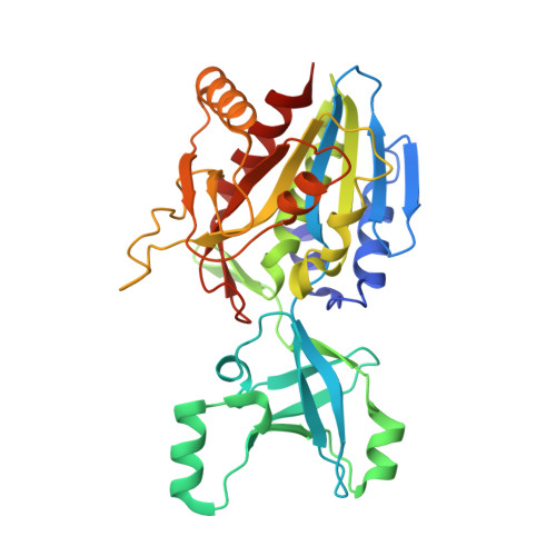
    The M42 aminopeptidases are dinuclear aminopeptidases displaying a peculiar tetrahedron-shaped structure with 12 subunits. Their quaternary structure results from the self-assembly of six dimers controlled by their divalent metal ion cofactors. The oligomeric-state transition remains debated despite the structural characterization of several archaeal M42 aminopeptidases. The main bottleneck is the lack of dimer structures, hindering the understanding of structural changes occurring during the oligomerization process. We present the first dimer structure of an M42 aminopeptidase, TmPep1050 of Thermotoga maritima , along with the dodecamer structure. The comparison of both structures has allowed us to describe how the metal ion cofactors modulate the active-site fold and, subsequently, affect the interaction interface between dimers. A mutational study shows that the M1 site strictly controls dodecamer formation. The dodecamer structure of TmPep1050 also reveals that a part of the dimerization domain delimits the catalytic pocket and could participate in substrate binding.


  • Organizational Affiliation
    • Laboratory of Microbiology, Department of Molecular Biology, Université Libre de Bruxelles, rue des Professeurs Jeener et Brachet 12, B6041 Charleroi, Belgium rdutoit@ulb.ac.be.

Macromolecule Content 

  • Total Structure Weight: 147.36 kDa 
  • Atom Count: 12,367 
  • Modeled Residue Count: 1,324 
  • Deposited Residue Count: 1,324 
  • Unique protein chains: 1

Macromolecules

Find similar proteins by:|  3D Structure
Entity ID: 1
MoleculeChains  Sequence LengthOrganismDetailsImage
Aminopeptidase
A, B, C, D
331Thermotoga maritima MSB8Mutation(s): 0 
Gene Names: TM_1050THEMA_09110Tmari_1054
EC: 3.2.1.4
UniProt
Find proteins for Q9X0E0 (Thermotoga maritima (strain ATCC 43589 / DSM 3109 / JCM 10099 / NBRC 100826 / MSB8))
Explore Q9X0E0 
Go to UniProtKB:  Q9X0E0
Entity Groups
Sequence Clusters30% Identity50% Identity70% Identity90% Identity95% Identity100% Identity
UniProt GroupQ9X0E0
Sequence Annotations
Expand
Reference Sequence

Small Molecules

Ligands 4 Unique
IDChains Name / Formula / InChI Key2D Diagram3D Interactions
SO4

Query on SO4


Download:Ideal Coordinates CCD File
AA [auth C]
BA [auth C]
CA [auth C]
FA [auth D]
G [auth A]
AA [auth C],
BA [auth C],
CA [auth C],
FA [auth D],
G [auth A],
GA [auth D],
H [auth A],
HA [auth D],
I [auth A],
IA [auth D],
J [auth A],
JA [auth D],
K [auth A],
KA [auth D],
L [auth A],
O [auth B],
P [auth B],
Q [auth B],
R [auth B],
S [auth B],
T [auth B],
U [auth B],
V [auth B],
Y [auth C],
Z [auth C]
SULFATE ION
O4 S
QAOWNCQODCNURD-UHFFFAOYSA-L
ZN
(Subject of Investigation/LOI)

Query on ZN


Download:Ideal Coordinates CCD File
DA [auth D],
E [auth A],
M [auth B],
W [auth C]
ZINC ION
Zn
PTFCDOFLOPIGGS-UHFFFAOYSA-N
CO
(Subject of Investigation/LOI)

Query on CO


Download:Ideal Coordinates CCD File
EA [auth D],
F [auth A],
N [auth B],
X [auth C]
COBALT (II) ION
Co
XLJKHNWPARRRJB-UHFFFAOYSA-N
OH
(Subject of Investigation/LOI)

Query on OH


Download:Ideal Coordinates CCD File
LA [auth D]HYDROXIDE ION
H O
XLYOFNOQVPJJNP-UHFFFAOYSA-M

Experimental Data & Validation

Experimental Data

  • Method: X-RAY DIFFRACTION
  • Resolution: 1.70 Å
  • R-Value Free:  0.164 (Depositor), 0.162 (DCC) 
  • R-Value Work:  0.143 (Depositor), 0.141 (DCC) 
  • R-Value Observed: 0.144 (Depositor) 
Space Group: H 3
Unit Cell:
Length ( Å )Angle ( ˚ )
a = 131.152α = 90
b = 131.152β = 90
c = 285.61γ = 120
Software Package:
Software NamePurpose
XDSdata reduction
XSCALEdata scaling
PHASERphasing
PHENIXrefinement
PDB_EXTRACTdata extraction

Structure Validation

View Full Validation Report


Entry History 

Deposition Data

  • Released Date: 2019-10-23 
  • Deposition Author(s): Dutoit, R.

Revision History  (Full details and data files)

  • Version 1.0: 2019-10-23
    Type: Initial release
  • Version 1.1: 2019-11-20
    Changes: Database references, Derived calculations, Other
  • Version 1.2: 2019-12-04
    Changes: Database references
  • Version 1.3: 2024-01-24
    Changes: Data collection, Database references, Derived calculations, Refinement description