6NPR | pdb_00006npr

Crystal structure of H-2Dd with C84-C139 disulfide in complex with gp120 derived peptide P18-I10


Experimental Data Snapshot

  • Method: X-RAY DIFFRACTION
  • Resolution: 2.37 Å
  • R-Value Free: 
    0.251 (Depositor), 0.257 (DCC) 
  • R-Value Work: 
    0.178 (Depositor), 0.186 (DCC) 
  • R-Value Observed: 
    0.182 (Depositor) 

Starting Model: experimental
View more details

wwPDB Validation 3D Report Full Report

Validation slider image for 6NPR

This is version 1.2 of the entry. See complete history

Literature

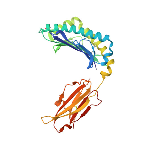
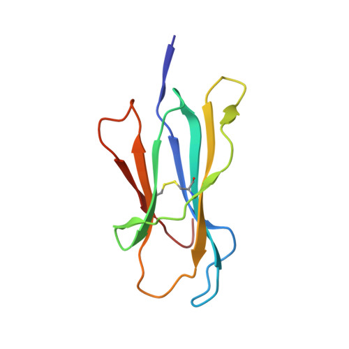
Molecular determinants of chaperone interactions on MHC-I for folding and antigen repertoire selection.

McShan, A.C.Devlin, C.A.Overall, S.A.Park, J.Toor, J.S.Moschidi, D.Flores-Solis, D.Choi, H.Tripathi, S.Procko, E.Sgourakis, N.G.

(2019) Proc Natl Acad Sci U S A 116: 25602-25613

  • DOI: https://doi.org/10.1073/pnas.1915562116
  • Primary Citation Related Structures: 
    6NPR

  • PubMed Abstract: 

    The interplay between a highly polymorphic set of MHC-I alleles and molecular chaperones shapes the repertoire of peptide antigens displayed on the cell surface for T cell surveillance. Here, we demonstrate that the molecular chaperone TAP-binding protein related (TAPBPR) associates with a broad range of partially folded MHC-I species inside the cell. Bimolecular fluorescence complementation and deep mutational scanning reveal that TAPBPR recognition is polarized toward the α 2 domain of the peptide-binding groove, and depends on the formation of a conserved MHC-I disulfide epitope in the α 2 domain. Conversely, thermodynamic measurements of TAPBPR binding for a representative set of properly conformed, peptide-loaded molecules suggest a narrower MHC-I specificity range. Using solution NMR, we find that the extent of dynamics at "hotspot" surfaces confers TAPBPR recognition of a sparsely populated MHC-I state attained through a global conformational change. Consistently, restriction of MHC-I groove plasticity through the introduction of a disulfide bond between the α 1 2 helices abrogates TAPBPR binding, both in solution and on a cellular membrane, while intracellular binding is tolerant of many destabilizing MHC-I substitutions. Our data support parallel TAPBPR functions of 1) chaperoning unstable MHC-I molecules with broad allele-specificity at early stages of their folding process, and 2) editing the peptide cargo of properly conformed MHC-I molecules en route to the surface, which demonstrates a narrower specificity. Our results suggest that TAPBPR exploits localized structural adaptations, both near and distant to the peptide-binding groove, to selectively recognize discrete conformational states sampled by MHC-I alleles, toward editing the repertoire of displayed antigens.


  • Organizational Affiliation
    • Department of Chemistry and Biochemistry, University of California, Santa Cruz, CA 95064.

Macromolecule Content 

  • Total Structure Weight: 90.09 kDa 
  • Atom Count: 6,482 
  • Modeled Residue Count: 768 
  • Deposited Residue Count: 772 
  • Unique protein chains: 3

Macromolecules

Find similar proteins by:|  3D Structure
Entity ID: 1
MoleculeChains  Sequence LengthOrganismDetailsImage
H-2 class I histocompatibility antigen, D-D alpha chain
A, C
276Mus musculusMutation(s): 0 
Gene Names: H2-D1
UniProt
Find proteins for P01900 (Mus musculus)
Explore P01900 
Go to UniProtKB:  P01900
Entity Groups
Sequence Clusters30% Identity50% Identity70% Identity90% Identity95% Identity100% Identity
UniProt GroupP01900
Sequence Annotations
Expand
Reference Sequence
Find similar proteins by:|  3D Structure
Entity ID: 2
MoleculeChains  Sequence LengthOrganismDetailsImage
Beta-2-microglobulin
B, D
100Homo sapiensMutation(s): 0 
Gene Names: B2MCDABP0092HDCMA22P
UniProt & NIH Common Fund Data Resources
Find proteins for P61769 (Homo sapiens)
Explore P61769 
Go to UniProtKB:  P61769
PHAROS:  P61769
GTEx:  ENSG00000166710 
Entity Groups
Sequence Clusters30% Identity50% Identity70% Identity90% Identity95% Identity100% Identity
UniProt GroupP61769
Sequence Annotations
Expand
Reference Sequence
Find similar proteins by:  Sequence   |   3D Structure  
Entity ID: 3
MoleculeChains  Sequence LengthOrganismDetailsImage
ARG-GLY-PRO-GLY-ARG-ALA-PHE-VAL-THR-ILEE [auth P],
F [auth R]
10Human immunodeficiency virus 1Mutation(s): 0 
UniProt
Find proteins for P04578 (Human immunodeficiency virus type 1 group M subtype B (isolate HXB2))
Explore P04578 
Go to UniProtKB:  P04578
Entity Groups
Sequence Clusters30% Identity50% Identity70% Identity90% Identity95% Identity100% Identity
UniProt GroupP04578
Sequence Annotations
Expand
Reference Sequence

Experimental Data & Validation

Experimental Data

  • Method: X-RAY DIFFRACTION
  • Resolution: 2.37 Å
  • R-Value Free:  0.251 (Depositor), 0.257 (DCC) 
  • R-Value Work:  0.178 (Depositor), 0.186 (DCC) 
  • R-Value Observed: 0.182 (Depositor) 
Space Group: P 2 21 21
Unit Cell:
Length ( Å )Angle ( ˚ )
a = 66.99α = 90
b = 105.33β = 90
c = 117.27γ = 90
Software Package:
Software NamePurpose
PHENIXrefinement
MOSFLMdata reduction
Aimlessdata scaling
PHASERphasing

Structure Validation

View Full Validation Report


Entry History 

& Funding Information

Deposition Data


Funding OrganizationLocationGrant Number
National Institutes of Health/National Human Genome Research Institute (NIH/NHGRI)United States1R35GM125034-01
National Institutes of Health/National Institute Of Allergy and Infectious Diseases (NIH/NIAID)United States1R01AI143997

Revision History  (Full details and data files)

  • Version 1.0: 2020-01-22
    Type: Initial release
  • Version 1.1: 2023-10-11
    Changes: Data collection, Database references, Refinement description
  • Version 1.2: 2024-10-23
    Changes: Structure summary