6NP0

Cryo-EM structure of 5HT3A receptor in presence of granisetron


Experimental Data Snapshot

  • Method: ELECTRON MICROSCOPY
  • Resolution: 2.92 Å
  • Aggregation State: PARTICLE 
  • Reconstruction Method: SINGLE PARTICLE 

wwPDB Validation   3D Report Full Report


This is version 2.0 of the entry. See complete history


Literature

Molecular mechanism of setron-mediated inhibition of full-length 5-HT3Areceptor.

Basak, S.Gicheru, Y.Kapoor, A.Mayer, M.L.Filizola, M.Chakrapani, S.

(2019) Nat Commun 10: 3225-3225

  • DOI: https://doi.org/10.1038/s41467-019-11142-8
  • Primary Citation of Related Structures:  
    6NP0

  • PubMed Abstract: 

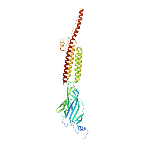
    Serotonin receptor (5-HT 3A R) is the most common therapeutic target to manage the nausea and vomiting during cancer therapies and in the treatment of irritable bowel syndrome. Setrons, a class of competitive antagonists, cause functional inhibition of 5-HT 3A R in the gastrointestinal tract and brainstem, acting as effective anti-emetic agents. Despite their prevalent use, the molecular mechanisms underlying setron binding and inhibition of 5-HT 3A R are not fully understood. Here, we present the structure of granisetron-bound full-length 5-HT 3A R solved by single-particle cryo-electron microscopy to 2.92 Å resolution. The reconstruction reveals the orientation of granisetron in the orthosteric site with unambiguous density for interacting sidechains. Molecular dynamics simulations and electrophysiology confirm the granisetron binding orientation and the residues central for ligand recognition. Comparison of granisetron-bound 5-HT 3A R with the apo and serotonin-bound structures, reveals key insights into the mechanism underlying 5-HT 3A R inhibition.


  • Organizational Affiliation

    Department of Physiology and Biophysics, Case Western Reserve University, Cleveland, OH, 44106-4970, USA.


Macromolecules
Find similar proteins by:  (by identity cutoff)  |  3D Structure
Entity ID: 1
MoleculeChains Sequence LengthOrganismDetailsImage
5-hydroxytryptamine receptor 3A
A, B, C, D, E
489Mus musculusMutation(s): 0 
Gene Names: Htr3a
Membrane Entity: Yes 
UniProt & NIH Common Fund Data Resources
Find proteins for P23979 (Mus musculus)
Explore P23979 
Go to UniProtKB:  P23979
IMPC:  MGI:96282
Entity Groups  
Sequence Clusters30% Identity50% Identity70% Identity90% Identity95% Identity100% Identity
UniProt GroupP23979
Sequence Annotations
Expand
  • Reference Sequence
Oligosaccharides

Help

Entity ID: 2
MoleculeChains Length2D Diagram Glycosylation3D Interactions
beta-D-mannopyranose-(1-4)-2-acetamido-2-deoxy-beta-D-glucopyranose-(1-4)-2-acetamido-2-deoxy-beta-D-glucopyranose
F, H, J, L, N
3N/A
Glycosylation Resources
GlyTouCan:  G15407YE
GlyCosmos:  G15407YE
GlyGen:  G15407YE
Entity ID: 3
MoleculeChains Length2D Diagram Glycosylation3D Interactions
2-acetamido-2-deoxy-beta-D-glucopyranose-(1-4)-2-acetamido-2-deoxy-beta-D-glucopyranose
G, I, K, M, O
2N-Glycosylation
Glycosylation Resources
GlyTouCan:  G42666HT
GlyCosmos:  G42666HT
GlyGen:  G42666HT
Small Molecules
Ligands 3 Unique
IDChains Name / Formula / InChI Key2D Diagram3D Interactions
CWB
Query on CWB

Download Ideal Coordinates CCD File 
BA [auth D],
CA [auth D],
R [auth A],
V [auth B],
Y [auth C]
1-methyl-N-[(1R,5S)-9-methyl-9-azabicyclo[3.3.1]nonan-3-yl]indazole-3-carboxamide
C18 H24 N4 O
MFWNKCLOYSRHCJ-BTTYYORXSA-N
NAG
Query on NAG

Download Ideal Coordinates CCD File 
AA [auth D]
DA [auth E]
EA [auth E]
P [auth A]
Q [auth A]
AA [auth D],
DA [auth E],
EA [auth E],
P [auth A],
Q [auth A],
T [auth B],
U [auth B],
W [auth C],
X [auth C],
Z [auth D]
2-acetamido-2-deoxy-beta-D-glucopyranose
C8 H15 N O6
OVRNDRQMDRJTHS-FMDGEEDCSA-N
CL
Query on CL

Download Ideal Coordinates CCD File 
S [auth A]CHLORIDE ION
Cl
VEXZGXHMUGYJMC-UHFFFAOYSA-M
Binding Affinity Annotations 
IDSourceBinding Affinity
CWB BindingDB:  6NP0 Ki: min: 0.25, max: 1000 (nM) from 12 assay(s)
Kd: min: 0.2, max: 1.4 (nM) from 4 assay(s)
IC50: 1760 (nM) from 1 assay(s)
EC50: min: 320, max: 500 (nM) from 2 assay(s)
Experimental Data & Validation

Experimental Data

  • Method: ELECTRON MICROSCOPY
  • Resolution: 2.92 Å
  • Aggregation State: PARTICLE 
  • Reconstruction Method: SINGLE PARTICLE 
EM Software:
TaskSoftware PackageVersion
RECONSTRUCTIONRELION3.0
MODEL REFINEMENTPHENIX1.13-2998

Structure Validation

View Full Validation Report


Entry History & Funding Information

Deposition Data


Funding OrganizationLocationGrant Number
National Institutes of Health/National Institute of General Medical Sciences (NIH/NIGMS)United States1R01GM108921
National Institutes of Health/National Institute of General Medical Sciences (NIH/NIGMS)United States3R01GM108921-03S1
American Heart AssociationUnited States17POST33671152

Revision History  (Full details and data files)

  • Version 1.0: 2019-07-24
    Type: Initial release
  • Version 1.1: 2019-07-31
    Changes: Data collection, Database references
  • Version 1.2: 2020-01-08
    Changes: Author supporting evidence
  • Version 2.0: 2020-07-29
    Type: Remediation
    Reason: Carbohydrate remediation
    Changes: Advisory, Atomic model, Data collection, Derived calculations, Structure summary