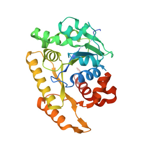
6NOP

Structure of Cyanothece McdA(D38A)-ATP complex


Experimental Data Snapshot

  • Method: X-RAY DIFFRACTION
  • Resolution: 1.70 Å
  • R-Value Free: 0.204 
  • R-Value Work: 0.182 
  • R-Value Observed: 0.183 

Starting Model: experimental
View more details

wwPDB Validation   3D Report Full Report


Ligand Structure Quality Assessment 


This is version 1.3 of the entry. See complete history


Literature

Structures of maintenance of carboxysome distribution Walker-box McdA and McdB adaptor homologs.

Schumacher, M.A.Henderson, M.Zhang, H.

(2019) Nucleic Acids Res 47: 5950-5962

  • DOI: https://doi.org/10.1093/nar/gkz314
  • Primary Citation of Related Structures:  
    6NON, 6NOO, 6NOP, 6NOY

  • PubMed Abstract: 

    Carboxysomes, protein-coated organelles in cyanobacteria, are important in global carbon fixation. However, these organelles are present at low copy in each cell and hence must be segregated to ensure transmission from one generation to the next. Recent studies revealed that a DNA partition-like ParA-ParB system mediates carboxysome maintenance, called McdA-McdB. Here, we describe the first McdA and McdB homolog structures. McdA is similar to partition ParA Walker-box proteins, but lacks the P-loop signature lysine involved in ATP binding. Strikingly, a McdA-ATP structure shows that a lysine distant from the P-loop and conserved in McdA homologs, enables ATP-dependent nucleotide sandwich dimer formation. Similar to partition ParA proteins this ATP-bound form binds nonspecific-DNA. McdB, which we show directly binds McdA, harbors a unique fold and appears to form higher-order oligomers like partition ParB proteins. Thus, our data reveal a new signature motif that enables McdA dimer formation and indicates that, similar to DNA segregation, carboxysome maintenance systems employ Walker-box proteins as DNA-binding motors while McdB proteins form higher order oligomers, which could function as adaptors to link carboxysomes and provide for stable transport by the McdA proteins.


  • Organizational Affiliation

    Department of Biochemistry, Duke University School of Medicine, Durham, NC 27710, USA.


Macromolecules
Find similar proteins by:  (by identity cutoff)  |  3D Structure
Entity ID: 1
MoleculeChains Sequence LengthOrganismDetailsImage
Cobyrinic acid ac-diamide synthaseA [auth B],
B [auth A]
278CyanotheceMutation(s): 1 
Gene Names: PCC7424_5529
EC: 3.6.4
UniProt
Find proteins for B7KMS4 (Gloeothece citriformis (strain PCC 7424))
Explore B7KMS4 
Go to UniProtKB:  B7KMS4
Entity Groups  
Sequence Clusters30% Identity50% Identity70% Identity90% Identity95% Identity100% Identity
UniProt GroupB7KMS4
Sequence Annotations
Expand
  • Reference Sequence
Experimental Data & Validation

Experimental Data

  • Method: X-RAY DIFFRACTION
  • Resolution: 1.70 Å
  • R-Value Free: 0.204 
  • R-Value Work: 0.182 
  • R-Value Observed: 0.183 
  • Space Group: P 31 2 1
Unit Cell:
Length ( Å )Angle ( ˚ )
a = 115.658α = 90
b = 115.658β = 90
c = 76.648γ = 120
Software Package:
Software NamePurpose
MOSFLMdata reduction
SCALAdata scaling
MOLREPphasing
PHENIXrefinement
PDB_EXTRACTdata extraction

Structure Validation

View Full Validation Report


Ligand Structure Quality Assessment 


Entry History 

Deposition Data

Revision History  (Full details and data files)

  • Version 1.0: 2019-04-24
    Type: Initial release
  • Version 1.1: 2019-06-05
    Changes: Data collection, Database references
  • Version 1.2: 2019-06-26
    Changes: Data collection, Database references
  • Version 1.3: 2023-10-11
    Changes: Data collection, Database references, Derived calculations, Refinement description