6NN4 | pdb_00006nn4

The structure of human liver pyruvate kinase, hLPYK-D499N, in complex with Fru-1,6-BP


Experimental Data Snapshot

  • Method: X-RAY DIFFRACTION
  • Resolution: 2.15 Å
  • R-Value Free: 
    0.253 (Depositor), 0.253 (DCC) 
  • R-Value Work: 
    0.230 (Depositor), 0.231 (DCC) 
  • R-Value Observed: 
    0.231 (Depositor) 

Starting Model: experimental
View more details

wwPDB Validation 3D Report Full Report

Validation slider image for 6NN4

Ligand Structure Quality Assessment 


This is version 1.4 of the entry. See complete history

Literature

Changes in the allosteric site of human liver pyruvate kinase upon activator binding include the breakage of an intersubunit cation-pi bond.

McFarlane, J.S.Ronnebaum, T.A.Meneely, K.M.Chilton, A.Fenton, A.W.Lamb, A.L.

(2019) Acta Crystallogr F Struct Biol Commun 75: 461-469

  • DOI: https://doi.org/10.1107/S2053230X19007209
  • Primary Citation Related Structures: 
    6NN4, 6NN5, 6NN7, 6NN8

  • PubMed Abstract: 

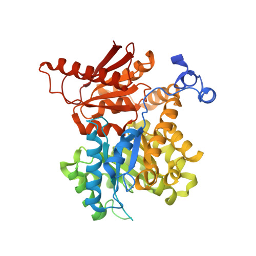
    Human liver pyruvate kinase (hLPYK) converts phosphoenolpyruvate to pyruvate in the final step of glycolysis. hLPYK is allosterically activated by fructose-1,6-bisphosphate (Fru-1,6-BP). The allosteric site, as defined by previous structural studies, is located in domain C between the phosphate-binding loop (residues 444-449) and the allosteric loop (residues 527-533). In this study, the X-ray crystal structures of four hLPYK variants were solved to make structural correlations with existing functional data. The variants are D499N, W527H, Δ529/S531G (called GGG here) and S531E. The results revealed a conformational toggle between the open and closed positions of the allosteric loop. In the absence of Fru-1,6-BP the open position is stabilized, in part, by a cation-π bond between Trp527 and Arg538' (from an adjacent monomer). In the S531E variant glutamate binds in place of the 6'-phosphate of Fru-1,6-BP in the allosteric site, leading to partial allosteric activation. Finally, the structure of the D499N mutant does not provide structural evidence for the previously observed allosteric activation of the D499N variant.


  • Organizational Affiliation
    • Department of Molecular Biosciences, University of Kansas, 1200 Sunnyside, Lawrence, KS 66045, USA.

Macromolecule Content 

  • Total Structure Weight: 236.72 kDa 
  • Atom Count: 13,236 
  • Modeled Residue Count: 1,665 
  • Deposited Residue Count: 2,172 
  • Unique protein chains: 1

Macromolecules

Find similar proteins by:|  3D Structure
Entity ID: 1
MoleculeChains  Sequence LengthOrganismDetailsImage
Pyruvate kinase PKLR
A, B, C, D
543Homo sapiensMutation(s): 1 
Gene Names: PKLRPK1PKL
EC: 2.7.1.40
UniProt & NIH Common Fund Data Resources
Find proteins for P30613 (Homo sapiens)
Explore P30613 
Go to UniProtKB:  P30613
PHAROS:  P30613
GTEx:  ENSG00000143627 
Entity Groups
Sequence Clusters30% Identity50% Identity70% Identity90% Identity95% Identity100% Identity
UniProt GroupP30613
Sequence Annotations
Expand
Reference Sequence

Small Molecules

Ligands 3 Unique
IDChains Name / Formula / InChI Key2D Diagram3D Interactions
FBP
(Subject of Investigation/LOI)

Query on FBP


Download:Ideal Coordinates CCD File
E [auth A],
I [auth B],
L [auth C],
O [auth D]
1,6-di-O-phosphono-beta-D-fructofuranose
C6 H14 O12 P2
RNBGYGVWRKECFJ-ARQDHWQXSA-N
PEP

Query on PEP


Download:Ideal Coordinates CCD File
H [auth A],
K [auth B],
N [auth C],
S [auth D]
PHOSPHOENOLPYRUVATE
C3 H5 O6 P
DTBNBXWJWCWCIK-UHFFFAOYSA-N
EDO

Query on EDO


Download:Ideal Coordinates CCD File
F [auth A]
G [auth A]
J [auth B]
M [auth C]
P [auth D]
F [auth A],
G [auth A],
J [auth B],
M [auth C],
P [auth D],
Q [auth D],
R [auth D]
1,2-ETHANEDIOL
C2 H6 O2
LYCAIKOWRPUZTN-UHFFFAOYSA-N

Experimental Data & Validation

Experimental Data

  • Method: X-RAY DIFFRACTION
  • Resolution: 2.15 Å
  • R-Value Free:  0.253 (Depositor), 0.253 (DCC) 
  • R-Value Work:  0.230 (Depositor), 0.231 (DCC) 
  • R-Value Observed: 0.231 (Depositor) 
Space Group: P 21 21 2
Unit Cell:
Length ( Å )Angle ( ˚ )
a = 120.572α = 90
b = 204.662β = 90
c = 112.334γ = 90
Software Package:
Software NamePurpose
PHENIXrefinement
XDSdata reduction
XDSdata scaling
PHASERphasing

Structure Validation

View Full Validation Report


Ligand Structure Quality Assessment 


Entry History 

& Funding Information

Deposition Data


Funding OrganizationLocationGrant Number
National Institutes of Health/National Human Genome Research Institute (NIH/NHGRI)United StatesR01GM115340
National Institutes of Health/National Human Genome Research Institute (NIH/NHGRI)United StatesR01GM127655
National Science Foundation (NSF, United States)United StatesCHE1403293
National Institutes of Health/National Human Genome Research Institute (NIH/NHGRI)United StatesT32GM08545
American Heart AssociationUnited StatesPRE33960374

Revision History  (Full details and data files)

  • Version 1.0: 2019-06-19
    Type: Initial release
  • Version 1.1: 2019-11-13
    Changes: Database references
  • Version 1.2: 2019-11-27
    Changes: Author supporting evidence
  • Version 1.3: 2020-07-29
    Type: Remediation
    Reason: Carbohydrate remediation
    Changes: Data collection, Derived calculations, Structure summary
  • Version 1.4: 2023-10-11
    Changes: Data collection, Database references, Refinement description, Structure summary