6NK5 | pdb_00006nk5

Electron Cryo-Microscopy Of Chikungunya VLP


Experimental Data Snapshot

  • Method: ELECTRON MICROSCOPY
  • Resolution: 4.16 Å
  • Aggregation State: PARTICLE 
  • Reconstruction Method: SINGLE PARTICLE 

Starting Models: experimental
View more details

wwPDB Validation 3D Report Full Report

Validation slider image for 6NK5

This is version 1.5 of the entry. See complete history. 

Literature

Cryo-EM Structure of Chikungunya Virus in Complex with the Mxra8 Receptor.

Basore, K., Kim, A.S., Nelson, C.A., Zhang, R., Smith, B.K., Uranga, C., Vang, L., Cheng, M., Gross, M.L., Smith, J., Diamond, M.S., Fremont, D.H.

(2019) Cell 177: 1725

  • DOI: https://doi.org/10.1016/j.cell.2019.04.006
  • Primary Citation Related Structures: 
    6NK3, 6NK5, 6NK6, 6NK7

  • PubMed Abstract: 

    Mxra8 is a receptor for multiple arthritogenic alphaviruses that cause debilitating acute and chronic musculoskeletal disease in humans. Herein, we present a 2.2 Å resolution X-ray crystal structure of Mxra8 and 4 to 5 Å resolution cryo-electron microscopy reconstructions of Mxra8 bound to chikungunya (CHIKV) virus-like particles and infectious virus. The Mxra8 ectodomain contains two strand-swapped Ig-like domains oriented in a unique disulfide-linked head-to-head arrangement. Mxra8 binds by wedging into a cleft created by two adjacent CHIKV E2-E1 heterodimers in one trimeric spike and engaging a neighboring spike. Two binding modes are observed with the fully mature VLP, with one Mxra8 binding with unique contacts. Only the high-affinity binding mode was observed in the complex with infectious CHIKV, as viral maturation and E3 occupancy appear to influence receptor binding-site usage. Our studies provide insight into how Mxra8 binds CHIKV and creates a path for developing alphavirus entry inhibitors.


  • Organizational Affiliation: 
    • Department of Pathology & Immunology, Washington University School of Medicine, Saint Louis, MO 63110, USA.

Macromolecule Content 

  • Total Structure Weight: 444.42 kDa 
  • Atom Count: 31,130 
  • Modeled Residue Count: 4,036 
  • Deposited Residue Count: 4,036 
  • Unique protein chains: 3

Macromolecules

Find similar proteins by:|  3D Structure
Entity ID: 1
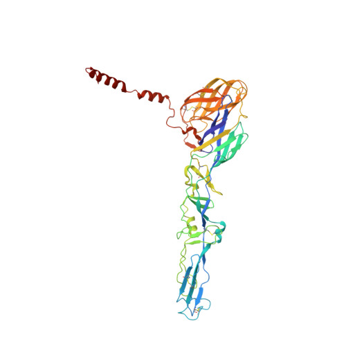
MoleculeChains  Sequence LengthOrganismDetailsImage
E1 glycoproteinA,
D [auth B],
G [auth C],
J [auth D]
439Chikungunya virus strain Senegal 37997Mutation(s): 0 
Membrane Entity: Yes 
UniProt
Find proteins for Q5XXP3 (Chikungunya virus (strain 37997))
Explore Q5XXP3 
Go to UniProtKB:  Q5XXP3
Entity Groups
Sequence Clusters30% Identity50% Identity70% Identity90% Identity95% Identity100% Identity
UniProt GroupQ5XXP3
Glycosylation
Glycosylation Sites: 1
Sequence Annotations
Expand
Reference Sequence
Find similar proteins by:|  3D Structure
Entity ID: 2
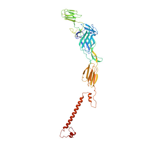
MoleculeChains  Sequence LengthOrganismDetailsImage
E2 glycoproteinB [auth E],
E [auth F],
H [auth G],
K [auth H]
419Chikungunya virus strain Senegal 37997Mutation(s): 0 
Membrane Entity: Yes 
UniProt
Find proteins for Q5XXP3 (Chikungunya virus (strain 37997))
Explore Q5XXP3 
Go to UniProtKB:  Q5XXP3
Entity Groups
Sequence Clusters30% Identity50% Identity70% Identity90% Identity95% Identity100% Identity
UniProt GroupQ5XXP3
Glycosylation
Glycosylation Sites: 1
Sequence Annotations
Expand
Reference Sequence
Find similar proteins by:|  3D Structure
Entity ID: 3
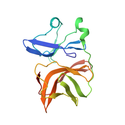
MoleculeChains  Sequence LengthOrganismDetailsImage
Capsid proteinC [auth I],
F [auth J],
I [auth K],
L
151Chikungunya virus strain Senegal 37997Mutation(s): 0 
Membrane Entity: Yes 
UniProt
Find proteins for Q5XXP3 (Chikungunya virus (strain 37997))
Explore Q5XXP3 
Go to UniProtKB:  Q5XXP3
Entity Groups
Sequence Clusters30% Identity50% Identity70% Identity90% Identity95% Identity100% Identity
UniProt GroupQ5XXP3
Sequence Annotations
Expand
Reference Sequence

Experimental Data & Validation

Experimental Data

  • Method: ELECTRON MICROSCOPY
  • Resolution: 4.16 Å
  • Aggregation State: PARTICLE 
  • Reconstruction Method: SINGLE PARTICLE 
EM Software:
TaskSoftware PackageVersion
RECONSTRUCTIONcisTEM1.0.0-beta
MODEL REFINEMENTPHENIX1.14

Structure Validation

View Full Validation Report


Entry History 

& Funding Information

Deposition Data


Funding OrganizationLocationGrant Number
National Institutes of Health/National Institute Of Allergy and Infectious Diseases (NIH/NIAID)United StatesHHSN272201700060C

Revision History  (Full details and data files)

  • Version 1.0: 2019-05-22
    Type: Initial release
  • Version 1.1: 2019-05-29
    Changes: Data collection, Database references
  • Version 1.2: 2019-06-26
    Changes: Data collection, Database references
  • Version 1.3: 2019-12-18
    Changes: Author supporting evidence
  • Version 1.4: 2020-07-29
    Type: Remediation
    Reason: Carbohydrate remediation
    Changes: Data collection, Derived calculations, Structure summary
  • Version 1.5: 2024-11-06
    Changes: Data collection, Database references, Derived calculations, Refinement description, Structure summary