6NEX | pdb_00006nex

Fab fragment of anti-cocaine antibody h2E2


Experimental Data Snapshot

  • Method: X-RAY DIFFRACTION
  • Resolution: 2.15 Å
  • R-Value Free: 
    0.254 (Depositor), 0.249 (DCC) 
  • R-Value Work: 
    0.214 (Depositor) 
  • R-Value Observed: 
    0.216 (Depositor) 

Starting Model: experimental
View more details

wwPDB Validation 3D Report Full Report

Validation slider image for 6NEX

This is version 2.1 of the entry. See complete history

Literature

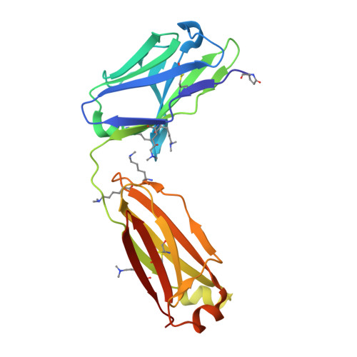
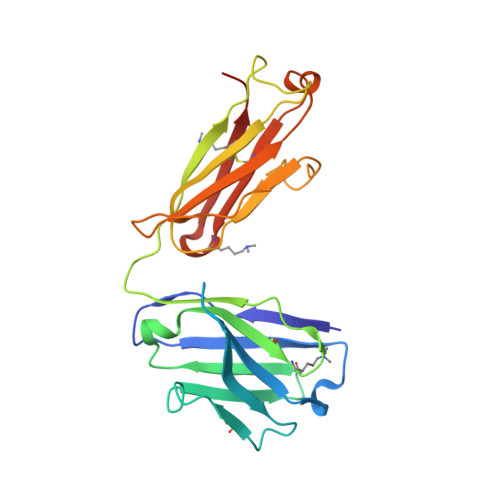
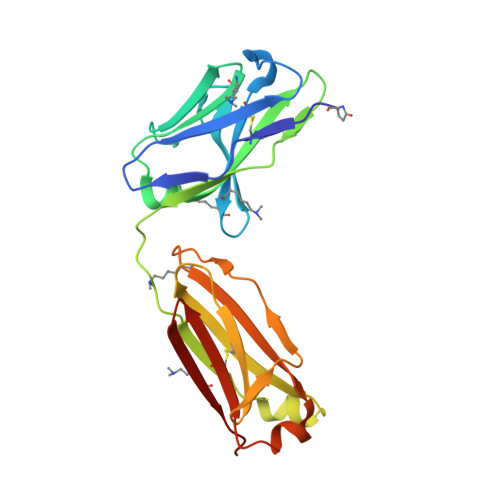
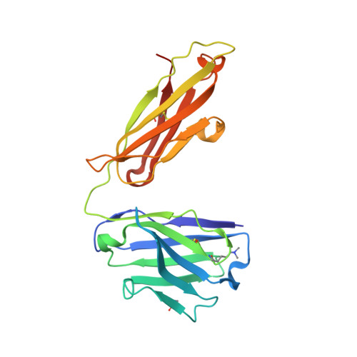
Structural analysis of free and liganded forms of the Fab fragment of a high-affinity anti-cocaine antibody, h2E2.

Tan, K.Zhou, M.Ahrendt, A.J.Duke, N.E.C.Tabaja, N.Ball, W.J.Kirley, T.L.Norman, A.B.Joachimiak, A.Schiffer, M.Wilton, R.Pokkuluri, P.R.

(2019) Acta Crystallogr F Struct Biol Commun 75: 697-706

  • DOI: https://doi.org/10.1107/S2053230X19013608
  • Primary Citation Related Structures: 
    6NEX, 6NFN

  • PubMed Abstract: 

    A high-affinity anti-cocaine monoclonal antibody, designated h2E2, is entering phase 1 clinical trials for cocaine abuse therapy. To gain insight into the molecular details of its structure that are important for binding cocaine and cocaine metabolites, the Fab fragment was generated and crystallized with and without ligand. Structures of the unliganded Fab and the Fab fragment bound to benzoylecgonine were determined, and were compared with each other and with other crystallized anti-cocaine antibodies. The affinity of the h2E2 antibody for cocaine is 4 nM, while that of the cocaine metabolite benzoylecgonine is 20 nM. Both are higher than the reported affinity for cocaine of the two previously crystallized anti-cocaine antibodies. Consistent with cocaine fluorescent quenching binding studies for the h2E2 mAb, four aromatic residues in the CDR regions of the Fab (TyrL32, TyrL96, TrpL91 and TrpH33) were found to be involved in ligand binding. The aromatic side chains surround and trap the tropane moiety of the ligand in the complex structure, forming significant van der Waals interactions which may account for the higher affinity observed for the h2E2 antibody. A water molecule mediates hydrogen bonding between the antibody and the carbonyl group of the benzoyl ester. The affinity of binding to h2E2 of benzoylecgonine differs only by a factor of five compared with that of cocaine; therefore, it is suggested that h2E2 would bind cocaine in the same way as observed in the Fab-benzoylecgonine complex, with minor rearrangements of some hypervariable segments of the antibody.


  • Organizational Affiliation
    • Structural Biology Center, X-ray Science Division, Argonne National Laboratory, Lemont, IL 60439, USA.

Macromolecule Content 

  • Total Structure Weight: 92.64 kDa 
  • Atom Count: 6,486 
  • Modeled Residue Count: 851 
  • Deposited Residue Count: 866 
  • Unique protein chains: 4

Macromolecules

Find similar proteins by:|  3D Structure
Entity ID: 1
MoleculeChains  Sequence LengthOrganismDetailsImage
Anitgen binding fragment light chainA [auth L]215Mus musculusMutation(s): 0 
Find similar proteins by:|  3D Structure
Entity ID: 2
MoleculeChains  Sequence LengthOrganismDetailsImage
Antigen binding fragment heavy chainB [auth H]218Mus musculusMutation(s): 0 
Find similar proteins by:|  3D Structure
Entity ID: 3
MoleculeChains  Sequence LengthOrganismDetailsImage
Anitgen binding fragment light chainC [auth A]215Mus musculusMutation(s): 0 
Find similar proteins by:|  3D Structure
Entity ID: 4
MoleculeChains  Sequence LengthOrganismDetailsImage
Ighg proteinD [auth B]218Mus musculusMutation(s): 0 
Gene Names: IghgAU044919

Small Molecules

Ligands 3 Unique
IDChains Name / Formula / InChI Key2D Diagram3D Interactions
ACT

Query on ACT


Download:Ideal Coordinates CCD File
J [auth H]ACETATE ION
C2 H3 O2
QTBSBXVTEAMEQO-UHFFFAOYSA-M
FMT

Query on FMT


Download:Ideal Coordinates CCD File
G [auth L],
H [auth L],
I [auth L],
L [auth A]
FORMIC ACID
C H2 O2
BDAGIHXWWSANSR-UHFFFAOYSA-N
CL

Query on CL


Download:Ideal Coordinates CCD File
E [auth L],
F [auth L],
K [auth A]
CHLORIDE ION
Cl
VEXZGXHMUGYJMC-UHFFFAOYSA-M
Modified Residues  2 Unique
IDChains TypeFormula2D DiagramParent
MLY
Query on MLY
A [auth L]L-PEPTIDE LINKINGC8 H18 N2 O2LYS
PCA
Query on PCA
A [auth L]L-PEPTIDE LINKINGC5 H7 N O3GLN

Experimental Data & Validation

Experimental Data

  • Method: X-RAY DIFFRACTION
  • Resolution: 2.15 Å
  • R-Value Free:  0.254 (Depositor), 0.249 (DCC) 
  • R-Value Work:  0.214 (Depositor) 
  • R-Value Observed: 0.216 (Depositor) 
Space Group: C 1 2 1
Unit Cell:
Length ( Å )Angle ( ˚ )
a = 110.787α = 90
b = 128.435β = 125.258
c = 91.196γ = 90
Software Package:
Software NamePurpose
SBC-Collectdata collection
PHENIXrefinement
HKL-3000data reduction
HKL-3000data scaling
PHASERphasing

Structure Validation

View Full Validation Report


Entry History 

Deposition Data

Revision History  (Full details and data files)

  • Version 1.0: 2019-11-20
    Type: Initial release
  • Version 2.0: 2020-01-22
    Changes: Polymer sequence, Structure summary
  • Version 2.1: 2024-04-03
    Changes: Data collection, Database references, Refinement description