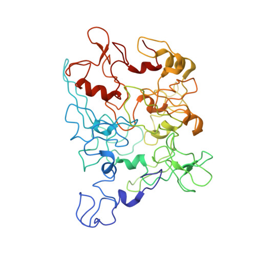
6NEF

Outer Membrane Cytochrome S Filament from Geobacter Sulfurreducens


Experimental Data Snapshot

  • Method: ELECTRON MICROSCOPY
  • Resolution: 3.40 Å
  • Aggregation State: FILAMENT 
  • Reconstruction Method: HELICAL 

wwPDB Validation   3D Report Full Report


This is version 2.1 of the entry. See complete history


Literature

Cryo-EM reveals the structural basis of long-range electron transport in a cytochrome-based bacterial nanowire.

Filman, D.J.Marino, S.F.Ward, J.E.Yang, L.Mester, Z.Bullitt, E.Lovley, D.R.Strauss, M.

(2019) Commun Biol 2: 219-219

  • DOI: https://doi.org/10.1038/s42003-019-0448-9
  • Primary Citation of Related Structures:  
    6NEF

  • PubMed Abstract: 

    Electrically conductive pili from Geobacter species, termed bacterial nanowires, are intensely studied for their biological significance and potential in the development of new materials. Using cryo-electron microscopy, we have characterized nanowires from conductive G. sulfurreducens pili preparations that are composed solely of head-to-tail stacked monomers of the six-heme C-type cytochrome OmcS. The unique fold of OmcS - closely wrapped around a continuous stack of hemes that can serve as an uninterrupted path for electron transport - generates a scaffold that supports the unbranched chain of hemes along the central axis of the filament. We present here, at 3.4 Å resolution, the structure of this cytochrome-based filament and discuss its possible role in long-range biological electron transport.


  • Organizational Affiliation

    1Department of Biological Chemistry and Molecular Pharmacology, Harvard Medical School, 240 Longwood Avenue, Boston, MA 02115 USA.


Macromolecules
Find similar proteins by:  (by identity cutoff)  |  3D Structure
Entity ID: 1
MoleculeChains Sequence LengthOrganismDetailsImage
C-type cytochrome OmcS407Geobacter sulfurreducens PCAMutation(s): 0 
UniProt
Find proteins for Q74A86 (Geobacter sulfurreducens (strain ATCC 51573 / DSM 12127 / PCA))
Explore Q74A86 
Go to UniProtKB:  Q74A86
Entity Groups  
Sequence Clusters30% Identity50% Identity70% Identity90% Identity95% Identity100% Identity
UniProt GroupQ74A86
Sequence Annotations
Expand
  • Reference Sequence
Experimental Data & Validation

Experimental Data

  • Method: ELECTRON MICROSCOPY
  • Resolution: 3.40 Å
  • Aggregation State: FILAMENT 
  • Reconstruction Method: HELICAL 
EM Software:
TaskSoftware PackageVersion
MODEL REFINEMENTREFMAC5.8.0238
MODEL REFINEMENTPHENIX

Structure Validation

View Full Validation Report


Entry History & Funding Information

Deposition Data


Funding OrganizationLocationGrant Number
Other governmentUnited StatesN00014-16-1-2526

Revision History  (Full details and data files)

  • Version 1.0: 2019-07-03
    Type: Initial release
  • Version 1.1: 2019-07-10
    Changes: Data collection, Database references
  • Version 2.0: 2019-09-11
    Changes: Atomic model, Data collection, Database references, Derived calculations, Non-polymer description, Source and taxonomy, Structure summary
  • Version 2.1: 2024-10-23
    Changes: Data collection, Database references, Derived calculations, Refinement description, Structure summary