


Structure of an HMG-CoA lyase from Acenitobacter baumannii in complex with coenzyme A and 3-methylmalate
Mayclin, S.J., Abendroth, J., Horanyi, P.S., Lorimer, D.D., Edwards, T.E.To be published.
Experimental Data Snapshot
Entity ID: 1 | |||||
|---|---|---|---|---|---|
| Molecule | Chains | Sequence Length | Organism | Details | Image |
| 3-hydroxy-3-methylglutaryl-CoA lyase | 320 | Acinetobacter baumannii | Mutation(s): 0 Gene Names: yngG_1, ABUW_2506, CBI29_01513, DV997_16665 EC: 4.1.3.4 | 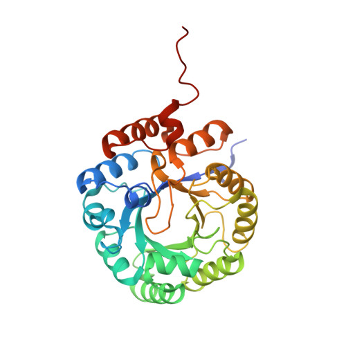 | |
UniProt | |||||
Find proteins for A0A0D5YK08 (Acinetobacter baumannii) Explore A0A0D5YK08 Go to UniProtKB: A0A0D5YK08 | |||||
Entity Groups | |||||
| Sequence Clusters | 30% Identity50% Identity70% Identity90% Identity95% Identity100% Identity | ||||
| UniProt Group | A0A0D5YK08 | ||||
Sequence AnnotationsExpand | |||||
Reference Sequence | |||||
| Ligands 5 Unique | |||||
|---|---|---|---|---|---|
| ID | Chains | Name / Formula / InChI Key | 2D Diagram | 3D Interactions | |
| COA Download:Ideal Coordinates CCD File | B [auth A] | COENZYME A C21 H36 N7 O16 P3 S RGJOEKWQDUBAIZ-IBOSZNHHSA-N |  | ||
| CIT Download:Ideal Coordinates CCD File | C [auth A] | CITRIC ACID C6 H8 O7 KRKNYBCHXYNGOX-UHFFFAOYSA-N |  | ||
| ZN Download:Ideal Coordinates CCD File | H [auth A] | ZINC ION Zn PTFCDOFLOPIGGS-UHFFFAOYSA-N |  | ||
| EDO Download:Ideal Coordinates CCD File | G [auth A] | 1,2-ETHANEDIOL C2 H6 O2 LYCAIKOWRPUZTN-UHFFFAOYSA-N |  | ||
| ACT Download:Ideal Coordinates CCD File | D [auth A], E [auth A], F [auth A] | ACETATE ION C2 H3 O2 QTBSBXVTEAMEQO-UHFFFAOYSA-M |  | ||
| Length ( Å ) | Angle ( ˚ ) |
|---|---|
| a = 91.46 | α = 90 |
| b = 91.46 | β = 90 |
| c = 78.85 | γ = 90 |
| Software Name | Purpose |
|---|---|
| PHENIX | refinement |
| XDS | data reduction |
| XSCALE | data scaling |
| PHASER | phasing |
| PDB_EXTRACT | data extraction |
| Coot | model building |
| MoRDa | phasing |


