6NCI | pdb_00006nci

Crystal structure of CDP-Chase: Vector data collection


Experimental Data Snapshot

  • Method: X-RAY DIFFRACTION
  • Resolution: 2.08 Å
  • R-Value Free: 
    0.223 (Depositor), 0.227 (DCC) 
  • R-Value Work: 
    0.171 (Depositor), 0.181 (DCC) 
  • R-Value Observed: 
    0.174 (Depositor) 

Starting Model: experimental
View more details

wwPDB Validation 3D Report Full Report

Validation slider image for 6NCI

Ligand Structure Quality Assessment 


This is version 2.1 of the entry. See complete history

Literature

Getting the Most Out of Your Crystals: Data Collection at the New High-Flux, Microfocus MX Beamlines at NSLS-II.

Miller, M.S.Maheshwari, S.Shi, W.Gao, Y.Chu, N.Soares, A.S.Cole, P.A.Amzel, L.M.Fuchs, M.R.Jakoncic, J.Gabelli, S.B.

(2019) Molecules 24

  • DOI: https://doi.org/10.3390/molecules24030496
  • Primary Citation Related Structures: 
    6NCH, 6NCI, 6NCK, 6NCT

  • PubMed Abstract: 

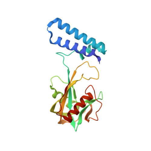
    Advances in synchrotron technology are changing the landscape of macromolecular crystallography. The two recently opened beamlines at NSLS-II-AMX and FMX-deliver high-flux microfocus beams that open new possibilities for crystallographic data collection. They are equipped with state-of-the-art experimental stations and automation to allow data collection on previously intractable crystals. Optimized data collection strategies allow users to tailor crystal positioning to optimally distribute the X-ray dose over its volume. Vector data collection allows the user to define a linear trajectory along a well diffracting volume of the crystal and perform rotational data collection while moving along the vector. This is particularly well suited to long, thin crystals. We describe vector data collection of three proteins-Akt1, PI3Kα, and CDP-Chase-to demonstrate its application and utility. For smaller crystals, we describe two methods for multicrystal data collection in a single loop, either manually selecting multiple centers (using H108A-PHM as an example), or "raster-collect", a more automated approach for a larger number of crystals (using CDP-Chase as an example).


  • Organizational Affiliation
    • Department of Oncology, Johns Hopkins University School of Medicine, Baltimore, MD 21205, USA. michelle.miller@jhmi.edu.

Macromolecule Content 

  • Total Structure Weight: 48.37 kDa 
  • Atom Count: 3,595 
  • Modeled Residue Count: 401 
  • Deposited Residue Count: 410 
  • Unique protein chains: 1

Macromolecules

Find similar proteins by:|  3D Structure
Entity ID: 1
MoleculeChains  Sequence LengthOrganismDetailsImage
Phosphohydrolase (MutT/nudix family protein)
A, B
205Bacillus cereus ATCC 14579Mutation(s): 0 
Gene Names: BC_2032
UniProt
Find proteins for Q81EE8 (Bacillus cereus (strain ATCC 14579 / DSM 31 / CCUG 7414 / JCM 2152 / NBRC 15305 / NCIMB 9373 / NCTC 2599 / NRRL B-3711))
Explore Q81EE8 
Go to UniProtKB:  Q81EE8
Entity Groups
Sequence Clusters30% Identity50% Identity70% Identity90% Identity95% Identity100% Identity
UniProt GroupQ81EE8
Sequence Annotations
Expand
Reference Sequence

Small Molecules

Ligands 4 Unique
IDChains Name / Formula / InChI Key2D Diagram3D Interactions
RB5
(Subject of Investigation/LOI)

Query on RB5


Download:Ideal Coordinates CCD File
I [auth B]D-ribose
C5 H10 O5
PYMYPHUHKUWMLA-LMVFSUKVSA-N
PEG

Query on PEG


Download:Ideal Coordinates CCD File
H [auth B]DI(HYDROXYETHYL)ETHER
C4 H10 O3
MTHSVFCYNBDYFN-UHFFFAOYSA-N
SO4

Query on SO4


Download:Ideal Coordinates CCD File
C [auth A]
D [auth A]
E [auth A]
F [auth A]
J [auth B]
C [auth A],
D [auth A],
E [auth A],
F [auth A],
J [auth B],
K [auth B]
SULFATE ION
O4 S
QAOWNCQODCNURD-UHFFFAOYSA-L
PO4
(Subject of Investigation/LOI)

Query on PO4


Download:Ideal Coordinates CCD File
G [auth A]PHOSPHATE ION
O4 P
NBIIXXVUZAFLBC-UHFFFAOYSA-K

Experimental Data & Validation

Experimental Data

  • Method: X-RAY DIFFRACTION
  • Resolution: 2.08 Å
  • R-Value Free:  0.223 (Depositor), 0.227 (DCC) 
  • R-Value Work:  0.171 (Depositor), 0.181 (DCC) 
  • R-Value Observed: 0.174 (Depositor) 
Space Group: P 21 21 21
Unit Cell:
Length ( Å )Angle ( ˚ )
a = 61.771α = 90
b = 67.008β = 90
c = 111.426γ = 90
Software Package:
Software NamePurpose
Aimlessdata scaling
MOLREPphasing
REFMACrefinement
PDB_EXTRACTdata extraction
XDSdata reduction

Structure Validation

View Full Validation Report


Ligand Structure Quality Assessment 


Entry History 

& Funding Information

Deposition Data


Funding OrganizationLocationGrant Number
National Institutes of Health/National Cancer Institute (NIH/NCI)United StatesCA062924

Revision History  (Full details and data files)

  • Version 1.0: 2019-02-06
    Type: Initial release
  • Version 1.1: 2019-11-13
    Changes: Database references
  • Version 1.2: 2019-12-04
    Changes: Author supporting evidence
  • Version 2.0: 2020-07-01
    Changes: Atomic model
  • Version 2.1: 2023-10-11
    Changes: Data collection, Database references, Refinement description