6NC4 | pdb_00006nc4

FtsY-NG high-resolution


Experimental Data Snapshot

  • Method: X-RAY DIFFRACTION
  • Resolution: 1.60 Å
  • R-Value Free: 
    0.189 (Depositor), 0.191 (DCC) 
  • R-Value Work: 
    0.159 (Depositor), 0.161 (DCC) 
  • R-Value Observed: 
    0.160 (Depositor) 

wwPDB Validation 3D Report Full Report

Validation slider image for 6NC4

Ligand Structure Quality Assessment 


This is version 1.3 of the entry. See complete history

Literature

Structural insights into the G-loop dynamics of E. coli FtsY NG domain.

Faoro, C.Ataide, S.F.

(2019) J Struct Biol 208: 107387-107387

  • DOI: https://doi.org/10.1016/j.jsb.2019.09.004
  • Primary Citation Related Structures: 
    6N5I, 6N5J, 6N6N, 6N9B, 6NC1, 6NC4

  • PubMed Abstract: 

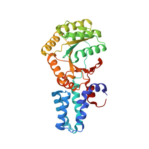
    The bacterial signal recognition particle (SRP) receptor, FtsY, participates with the SRP in co-translation targeting of proteins. Multiple crystal structures of the NG domain of E. coli FtsY NG have been determined at high-resolution (1.22-1.88 Å), in the nucleotide-free (apo) form as well as bound to GDP and non-hydrolysable GTP analogues. The combination of high-resolution and multiple solved structures of FtsY NG in different states revealed a new sensor-relay system of this unique GTPase receptor. A nucleotide sensing function of the P-loop assists FtsY NG in nucleotide-binding and contributes to modulate nucleotide binding properties in SRP GTPases. A reorganization of the other G-loops and the insertion binding domain (IBD) is observed only upon transition from a diphosphate to a triphosphate nucleotide. The role of a magnesium ion during the GDP and GTP-bound states has also been observed. The binding of magnesium in the nucleotide site causes the reorientation of the β- and γ- phosphate groups toward the jaws of the P-loop and stabilizes the binding of the nucleotide, creating a network of hydrogen and water-bridge interactions.


  • Organizational Affiliation
    • School of Life and Environmental Sciences, The University of Sydney, Sydney 2006, Australia.

Macromolecule Content 

  • Total Structure Weight: 68.09 kDa 
  • Atom Count: 5,268 
  • Modeled Residue Count: 606 
  • Deposited Residue Count: 606 
  • Unique protein chains: 1

Macromolecules

Find similar proteins by:|  3D Structure
Entity ID: 1
MoleculeChains  Sequence LengthOrganismDetailsImage
Signal recognition particle receptor FtsY
A, B
303Escherichia coli K-12Mutation(s): 0 
Gene Names: ftsYb3464JW3429
EC: 3.6.5.4
UniProt
Find proteins for P10121 (Escherichia coli (strain K12))
Explore P10121 
Go to UniProtKB:  P10121
Entity Groups
Sequence Clusters30% Identity50% Identity70% Identity90% Identity95% Identity100% Identity
UniProt GroupP10121
Sequence Annotations
Expand
Reference Sequence

Small Molecules

Ligands 5 Unique
IDChains Name / Formula / InChI Key2D Diagram3D Interactions
GNP

Query on GNP


Download:Ideal Coordinates CCD File
C [auth A]PHOSPHOAMINOPHOSPHONIC ACID-GUANYLATE ESTER
C10 H17 N6 O13 P3
UQABYHGXWYXDTK-UUOKFMHZSA-N
GDP

Query on GDP


Download:Ideal Coordinates CCD File
L [auth B]GUANOSINE-5'-DIPHOSPHATE
C10 H15 N5 O11 P2
QGWNDRXFNXRZMB-UUOKFMHZSA-N
GOL

Query on GOL


Download:Ideal Coordinates CCD File
M [auth B]GLYCEROL
C3 H8 O3
PEDCQBHIVMGVHV-UHFFFAOYSA-N
ACT

Query on ACT


Download:Ideal Coordinates CCD File
D [auth A]
E [auth A]
N [auth B]
O [auth B]
P [auth B]
D [auth A],
E [auth A],
N [auth B],
O [auth B],
P [auth B],
Q [auth B],
R [auth B],
S [auth B]
ACETATE ION
C2 H3 O2
QTBSBXVTEAMEQO-UHFFFAOYSA-M
NH4

Query on NH4


Download:Ideal Coordinates CCD File
AA [auth B]
BA [auth B]
CA [auth B]
DA [auth B]
EA [auth B]
AA [auth B],
BA [auth B],
CA [auth B],
DA [auth B],
EA [auth B],
F [auth A],
FA [auth B],
G [auth A],
GA [auth B],
H [auth A],
I [auth A],
J [auth A],
K [auth A],
T [auth B],
U [auth B],
V [auth B],
W [auth B],
X [auth B],
Y [auth B],
Z [auth B]
AMMONIUM ION
H4 N
QGZKDVFQNNGYKY-UHFFFAOYSA-O

Experimental Data & Validation

Experimental Data

  • Method: X-RAY DIFFRACTION
  • Resolution: 1.60 Å
  • R-Value Free:  0.189 (Depositor), 0.191 (DCC) 
  • R-Value Work:  0.159 (Depositor), 0.161 (DCC) 
  • R-Value Observed: 0.160 (Depositor) 
Space Group: P 1 21 1
Unit Cell:
Length ( Å )Angle ( ˚ )
a = 34.817α = 90
b = 75.905β = 93.39
c = 108.403γ = 90
Software Package:
Software NamePurpose
PHENIXrefinement
XDSdata reduction
Aimlessdata scaling
PHASERphasing

Structure Validation

View Full Validation Report


Ligand Structure Quality Assessment 


Entry History 

Deposition Data

Revision History  (Full details and data files)

  • Version 1.0: 2019-10-23
    Type: Initial release
  • Version 1.1: 2019-12-04
    Changes: Database references
  • Version 1.2: 2020-03-18
    Changes: Database references
  • Version 1.3: 2024-03-13
    Changes: Data collection, Database references