6NAX | pdb_00006nax

Olfactomedin domain of mouse myocilin


Experimental Data Snapshot

  • Method: X-RAY DIFFRACTION
  • Resolution: 1.55 Å
  • R-Value Free: 
    0.178 (Depositor), 0.177 (DCC) 
  • R-Value Work: 
    0.158 (Depositor), 0.159 (DCC) 
  • R-Value Observed: 
    0.158 (Depositor) 

Starting Model: experimental
View more details

wwPDB Validation 3D Report Full Report

Validation slider image for 6NAX

This is version 1.5 of the entry. See complete history

Literature

Differential Misfolding Properties of Glaucoma-Associated Olfactomedin Domains from Humans and Mice.

Patterson-Orazem, A.C.Hill, S.E.Wang, Y.Dominic, I.M.Hall, C.K.Lieberman, R.L.

(2019) Biochemistry 58: 1718-1727

  • DOI: https://doi.org/10.1021/acs.biochem.8b01309
  • Primary Citation Related Structures: 
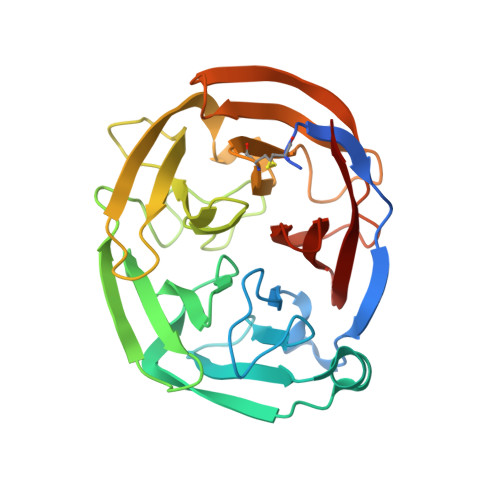
    6NAX

  • PubMed Abstract: 

    Mutations in myocilin, predominantly within its olfactomedin (OLF) domain, are causative for the heritable form of open angle glaucoma in humans. Surprisingly, mice expressing Tyr423His mutant myocilin, corresponding to a severe glaucoma-causing mutation (Tyr437His) in human subjects, exhibit a weak, if any, glaucoma phenotype. To address possible protein-level discrepancies between mouse and human OLFs, which might lead to this outcome, biophysical properties of mouse OLF were characterized for comparison with those of human OLF. The 1.55 Å resolution crystal structure of mouse OLF reveals an asymmetric 5-bladed β-propeller that is nearly indistinguishable from previous structures of human OLF. Wild-type and selected mutant mouse OLFs mirror thermal stabilities of their human OLF counterparts, including characteristic stabilization in the presence of calcium. Mouse OLF forms thioflavin T-positive aggregates with a similar end-point morphology as human OLF, but amyloid aggregation kinetic rates of mouse OLF are faster than human OLF. Simulations and experiments support the interpretation that kinetics of mouse OLF are faster because of a decreased charge repulsion arising from more neutral surface electrostatics. Taken together, phenotypic differences observed in mouse and human studies of mutant myocilin could be a function of aggregation kinetics rates, which would alter the lifetime of putatively toxic protofibrillar intermediates.


  • Organizational Affiliation
    • School of Chemistry & Biochemistry , Georgia Institute of Technology , Atlanta , Georgia 30332-0400 , United States.

Macromolecule Content 

  • Total Structure Weight: 62.99 kDa 
  • Atom Count: 4,857 
  • Modeled Residue Count: 518 
  • Deposited Residue Count: 554 
  • Unique protein chains: 1

Macromolecules

Find similar proteins by:|  3D Structure
Entity ID: 1
MoleculeChains  Sequence LengthOrganismDetailsImage
Myocilin
A, B
277Mus musculusMutation(s): 0 
Gene Names: MyocTigr
UniProt
Find proteins for O70624 (Mus musculus)
Explore O70624 
Go to UniProtKB:  O70624
Entity Groups
Sequence Clusters30% Identity50% Identity70% Identity90% Identity95% Identity100% Identity
UniProt GroupO70624
Sequence Annotations
Expand
Reference Sequence

Small Molecules

Ligands 4 Unique
IDChains Name / Formula / InChI Key2D Diagram3D Interactions
GOL

Query on GOL


Download:Ideal Coordinates CCD File
E [auth A],
F [auth A],
J [auth B],
K [auth B]
GLYCEROL
C3 H8 O3
PEDCQBHIVMGVHV-UHFFFAOYSA-N
CA

Query on CA


Download:Ideal Coordinates CCD File
C [auth A],
H [auth B]
CALCIUM ION
Ca
BHPQYMZQTOCNFJ-UHFFFAOYSA-N
K

Query on K


Download:Ideal Coordinates CCD File
G [auth A]POTASSIUM ION
K
NPYPAHLBTDXSSS-UHFFFAOYSA-N
NA

Query on NA


Download:Ideal Coordinates CCD File
D [auth A],
I [auth B]
SODIUM ION
Na
FKNQFGJONOIPTF-UHFFFAOYSA-N

Experimental Data & Validation

Experimental Data

  • Method: X-RAY DIFFRACTION
  • Resolution: 1.55 Å
  • R-Value Free:  0.178 (Depositor), 0.177 (DCC) 
  • R-Value Work:  0.158 (Depositor), 0.159 (DCC) 
  • R-Value Observed: 0.158 (Depositor) 
Space Group: P 4
Unit Cell:
Length ( Å )Angle ( ˚ )
a = 111.955α = 90
b = 111.955β = 90
c = 44.129γ = 90
Software Package:
Software NamePurpose
PHENIXrefinement
HKL-2000data reduction
HKL-2000data scaling
PHENIXphasing

Structure Validation

View Full Validation Report


Entry History 

& Funding Information

Deposition Data


Funding OrganizationLocationGrant Number
National Institutes of Health/National Eye Institute (NIH/NEI)United StatesR01EY021205
Other privateUnited StatesG2016027

Revision History  (Full details and data files)

  • Version 1.0: 2019-03-13
    Type: Initial release
  • Version 1.1: 2019-03-27
    Changes: Data collection, Database references
  • Version 1.2: 2019-04-10
    Changes: Data collection, Database references
  • Version 1.3: 2019-12-11
    Changes: Author supporting evidence
  • Version 1.4: 2023-10-11
    Changes: Data collection, Database references, Derived calculations, Refinement description
  • Version 1.5: 2024-10-16
    Changes: Structure summary