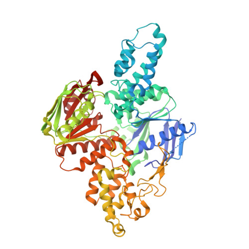
6N9L

Crystal structure of T. maritima UvrA d117-399 with ADP


Experimental Data Snapshot

  • Method: X-RAY DIFFRACTION
  • Resolution: 2.01 Å
  • R-Value Free: 0.201 
  • R-Value Work: 0.169 
  • R-Value Observed: 0.170 

Starting Model: experimental
View more details

wwPDB Validation   3D Report Full Report


Ligand Structure Quality Assessment 


This is version 1.3 of the entry. See complete history


Literature

The ATPase mechanism of UvrA2 reveals the distinct roles of proximal and distal ATPase sites in nucleotide excision repair.

Case, B.C.Hartley, S.Osuga, M.Jeruzalmi, D.Hingorani, M.M.

(2019) Nucleic Acids Res 47: 4136-4152

  • DOI: https://doi.org/10.1093/nar/gkz180
  • Primary Citation of Related Structures:  
    6N9L

  • PubMed Abstract: 

    The UvrA2 dimer finds lesions in DNA and initiates nucleotide excision repair. Each UvrA monomer contains two essential ATPase sites: proximal (P) and distal (D). The manner whereby their activities enable UvrA2 damage sensing and response remains to be clarified. We report three key findings from the first pre-steady state kinetic analysis of each site. Absent DNA, a P2ATP-D2ADP species accumulates when the low-affinity proximal sites bind ATP and enable rapid ATP hydrolysis and phosphate release by the high-affinity distal sites, and ADP release limits catalytic turnover. Native DNA stimulates ATP hydrolysis by all four sites, causing UvrA2 to transition through a different species, P2ADP-D2ADP. Lesion-containing DNA changes the mechanism again, suppressing ATP hydrolysis by the proximal sites while distal sites cycle through hydrolysis and ADP release, to populate proximal ATP-bound species, P2ATP-Dempty and P2ATP-D2ATP. Thus, damaged and native DNA trigger distinct ATPase site activities, which could explain why UvrA2 forms stable complexes with UvrB on damaged DNA compared with weaker, more dynamic complexes on native DNA. Such specific coupling between the DNA substrate and the ATPase mechanism of each site provides new insights into how UvrA2 utilizes ATP for lesion search, recognition and repair.


  • Organizational Affiliation

    Department of Molecular Biology and Biochemistry, Wesleyan University, Middletown, CT 06459, USA.


Macromolecules
Find similar proteins by:  (by identity cutoff)  |  3D Structure
Entity ID: 1
MoleculeChains Sequence LengthOrganismDetailsImage
UvrABC system protein A640Thermotoga maritimaMutation(s): 0 
Gene Names: uvrATM_0480
UniProt
Find proteins for Q9WYV0 (Thermotoga maritima (strain ATCC 43589 / DSM 3109 / JCM 10099 / NBRC 100826 / MSB8))
Explore Q9WYV0 
Go to UniProtKB:  Q9WYV0
Entity Groups  
Sequence Clusters30% Identity50% Identity70% Identity90% Identity95% Identity100% Identity
UniProt GroupQ9WYV0
Sequence Annotations
Expand
  • Reference Sequence
Experimental Data & Validation

Experimental Data

  • Method: X-RAY DIFFRACTION
  • Resolution: 2.01 Å
  • R-Value Free: 0.201 
  • R-Value Work: 0.169 
  • R-Value Observed: 0.170 
  • Space Group: C 1 2 1
Unit Cell:
Length ( Å )Angle ( ˚ )
a = 143.134α = 90
b = 81.379β = 125.3
c = 90.416γ = 90
Software Package:
Software NamePurpose
PHENIXrefinement
HKL-2000data reduction
HKL-2000data scaling
PHASERphasing

Structure Validation

View Full Validation Report


Ligand Structure Quality Assessment 


Entry History & Funding Information

Deposition Data


Funding OrganizationLocationGrant Number
National Science Foundation (NSF, United States)United StatesMCB# 1330528
National Institutes of Health/National Institute of General Medical Sciences (NIH/NIGMS)United StatesR15 GM114743

Revision History  (Full details and data files)

  • Version 1.0: 2019-05-01
    Type: Initial release
  • Version 1.1: 2019-07-03
    Changes: Data collection, Database references
  • Version 1.2: 2019-11-27
    Changes: Author supporting evidence
  • Version 1.3: 2023-10-11
    Changes: Data collection, Database references, Refinement description