6N96 | pdb_00006n96

Methylmalonyl-CoA decarboxylase in complex with 2-sulfonate-propionyl-oxa(dethia)-CoA


Experimental Data Snapshot

  • Method: X-RAY DIFFRACTION
  • Resolution: 1.70 Å
  • R-Value Free: 
    0.184 (Depositor), 0.192 (DCC) 
  • R-Value Work: 
    0.151 (Depositor), 0.163 (DCC) 
  • R-Value Observed: 
    0.152 (Depositor) 

Starting Model: experimental
View more details

wwPDB Validation 3D Report Full Report

Validation slider image for 6N96

Ligand Structure Quality Assessment 


This is version 1.1 of the entry. See complete history

Literature

Sulfonate/Nitro Bearing Methylmalonyl-Thioester Isosteres Applied to Methylmalonyl-CoA Decarboxylase Structure-Function Studies.

Stunkard, L.M.Dixon, A.D.Huth, T.J.Lohman, J.R.

(2019) J Am Chem Soc 141: 5121-5124

  • DOI: https://doi.org/10.1021/jacs.9b00650
  • Primary Citation Related Structures: 
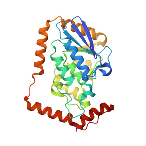
    6N92, 6N93, 6N94, 6N95, 6N96, 6N97

  • PubMed Abstract: 

    Malonyl-thioesters are reactive centers of malonyl-CoA and malonyl- S-acyl carrier protein, essential to fatty acid, polyketide and various specialized metabolite biosynthesis. Enzymes that create or use malonyl-thioesters spontaneously hydrolyze or decarboxylate reactants on the crystallographic time frame preventing determination of structure-function relationships. To address this problem, we have synthesized a panel of methylmalonyl-CoA analogs with the carboxylate represented by a sulfonate or nitro and the thioester retained or represented by an ester or amide. Structures of Escherichia coli methylmalonyl-CoA decarboxylase in complex with our analogs affords insight into substrate binding and the catalytic mechanism. Counterintuitively, the negatively charged sulfonate and nitronate functional groups of our analogs bind in an active site hydrophobic pocket. Upon decarboxylation the enolate intermediate is protonated by a histidine preventing CO 2 -enolate recombination, yielding propionyl-CoA. Activity assays support a histidine catalytic acid and reveal the enzyme displays significant hydrolysis activity. Our structures also provide insight into this hydrolysis activity. Our analogs inhibit decarboxylation/hydrolysis activity with low micromolar K i values. This study sets precedents for using malonyl-CoA analogs with carboxyate isosteres to study the complicated structure-function relationships of acyl-CoA carboxylases, trans-carboxytransferases, malonyltransferases and β-ketoacylsynthases.


  • Organizational Affiliation
    • Department of Biochemistry , Purdue University , West Lafayette , Indiana 47907 , United States.

Macromolecule Content 

  • Total Structure Weight: 187.64 kDa 
  • Atom Count: 15,313 
  • Modeled Residue Count: 1,560 
  • Deposited Residue Count: 1,566 
  • Unique protein chains: 1

Macromolecules

Find similar proteins by:|  3D Structure
Entity ID: 1
MoleculeChains  Sequence LengthOrganismDetailsImage
Methylmalonyl-CoA decarboxylase
A, B, C, D, E
A, B, C, D, E, F
261Escherichia coli K-12Mutation(s): 1 
Gene Names: scpBmmcDygfGb2919JW2886
EC: 4.1.1
UniProt
Find proteins for P52045 (Escherichia coli (strain K12))
Explore P52045 
Go to UniProtKB:  P52045
Entity Groups
Sequence Clusters30% Identity50% Identity70% Identity90% Identity95% Identity100% Identity
UniProt GroupP52045
Sequence Annotations
Expand
Reference Sequence

Small Molecules

Ligands 3 Unique
IDChains Name / Formula / InChI Key2D Diagram3D Interactions
SO5

Query on SO5


Download:Ideal Coordinates CCD File
H [auth A]
J [auth B]
L [auth C]
O [auth D]
R [auth E]
H [auth A],
J [auth B],
L [auth C],
O [auth D],
R [auth E],
T [auth F]
(2~{R})-1-[2-[3-[[(2~{R})-4-[[[(2~{R},3~{S},4~{R},5~{R})-5-(6-aminopurin-9-yl)-4-oxidanyl-3-phosphonooxy-oxolan-2-yl]methoxy-oxidanyl-phosphoryl]oxy-oxidanyl-phosphoryl]oxy-3,3-dimethyl-2-oxidanyl-butanoyl]amino]propanoylamino]ethoxy]-1-oxidanylidene-propane-2-sulfonic acid
C24 H40 N7 O21 P3 S
BEMUOEZGNHFKKE-AGCMQPJKSA-N
LCV

Query on LCV


Download:Ideal Coordinates CCD File
G [auth A]
I [auth B]
K [auth C]
M [auth C]
N [auth D]
G [auth A],
I [auth B],
K [auth C],
M [auth C],
N [auth D],
Q [auth E],
S [auth F],
U [auth F]
(2~{S})-1-[2-[3-[[(2~{R})-4-[[[(2~{R},3~{S},4~{R},5~{R})-5-(6-aminopurin-9-yl)-4-oxidanyl-3-phosphonooxy-oxolan-2-yl]methoxy-oxidanyl-phosphoryl]oxy-oxidanyl-phosphoryl]oxy-3,3-dimethyl-2-oxidanyl-butanoyl]amino]propanoylamino]ethoxy]-1-oxidanylidene-propane-2-sulfonic acid
C24 H40 N7 O21 P3 S
BEMUOEZGNHFKKE-IBNUZSNCSA-N
IMD

Query on IMD


Download:Ideal Coordinates CCD File
P [auth D]IMIDAZOLE
C3 H5 N2
RAXXELZNTBOGNW-UHFFFAOYSA-O

Experimental Data & Validation

Experimental Data

  • Method: X-RAY DIFFRACTION
  • Resolution: 1.70 Å
  • R-Value Free:  0.184 (Depositor), 0.192 (DCC) 
  • R-Value Work:  0.151 (Depositor), 0.163 (DCC) 
  • R-Value Observed: 0.152 (Depositor) 
Space Group: P 21 21 21
Unit Cell:
Length ( Å )Angle ( ˚ )
a = 87.148α = 90
b = 114.66β = 90
c = 192.705γ = 90
Software Package:
Software NamePurpose
REFMACrefinement
PDB_EXTRACTdata extraction
HKL-2000data reduction
HKL-2000data scaling
REFMACphasing

Structure Validation

View Full Validation Report


Ligand Structure Quality Assessment 


Entry History 

Deposition Data

Revision History  (Full details and data files)

  • Version 1.0: 2019-04-10
    Type: Initial release
  • Version 1.1: 2023-10-11
    Changes: Data collection, Database references, Refinement description