6N87

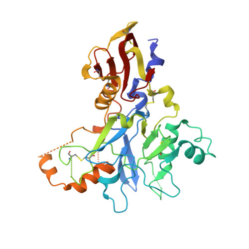
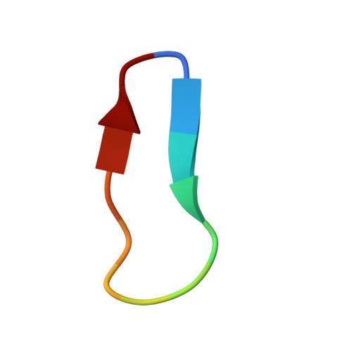
Plasmodium falciparum FVO apical membrane antigen 1 (AMA1) bound to MTSL spin-labelled cyclised RON2 peptide


Experimental Data Snapshot

  • Method: X-RAY DIFFRACTION
  • Resolution: 1.59 Å
  • R-Value Free: 0.188 
  • R-Value Work: 0.160 
  • R-Value Observed: 0.162 

Starting Model: experimental
View more details

wwPDB Validation   3D Report Full Report


Ligand Structure Quality Assessment 


This is version 1.4 of the entry. See complete history


Literature

Identification of the Binding Site of Apical Membrane Antigen 1 (AMA1) Inhibitors Using a Paramagnetic Probe.

Akter, M.Drinkwater, N.Devine, S.M.Drew, S.C.Krishnarjuna, B.Debono, C.O.Wang, G.Scanlon, M.J.Scammells, P.J.McGowan, S.MacRaild, C.A.Norton, R.S.

(2019) ChemMedChem 14: 603-612

  • DOI: https://doi.org/10.1002/cmdc.201800802
  • Primary Citation of Related Structures:  
    6N7Q, 6N87

  • PubMed Abstract: 

    Apical membrane antigen 1 (AMA1) is essential for the invasion of host cells by malaria parasites. Several small-molecule ligands have been shown to bind to a conserved hydrophobic cleft in Plasmodium falciparum AMA1. However, a lack of detailed structural information on the binding pose of these molecules has hindered their further optimisation as inhibitors. We have developed a spin-labelled peptide based on RON2, the native binding partner of AMA1, to probe the binding sites of compounds on PfAMA1. The crystal structure of this peptide bound to PfAMA1 shows that it binds at one end of the hydrophobic groove, leaving much of the binding site unoccupied and allowing fragment hits to bind without interference. In paramagnetic relaxation enhancement (PRE)-based NMR screening, the 1 H relaxation rates of compounds binding close to the probe were enhanced. Compounds experienced different degrees of PRE as a result of their different orientations relative to the spin label while bound to AMA1. Thus, PRE-derived distance constraints can be used to identify binding sites and guide further hit optimisation.


  • Organizational Affiliation

    Medicinal Chemistry, Monash Institute of Pharmaceutical Sciences, Monash University, Parkville, Victoria, 3052, Australia.


Macromolecules
Find similar proteins by:  (by identity cutoff)  |  3D Structure
Entity ID: 1
MoleculeChains Sequence LengthOrganismDetailsImage
Apical membrane antigen-1336Plasmodium falciparumMutation(s): 0 
Gene Names: ama-1
UniProt
Find proteins for Q1PBJ5 (Plasmodium falciparum)
Explore Q1PBJ5 
Go to UniProtKB:  Q1PBJ5
Entity Groups  
Sequence Clusters30% Identity50% Identity70% Identity90% Identity95% Identity100% Identity
UniProt GroupQ1PBJ5
Sequence Annotations
Expand
  • Reference Sequence

Find similar proteins by:  Sequence   |   3D Structure  

Entity ID: 2
MoleculeChains Sequence LengthOrganismDetailsImage
backbone-cyclised peptide bcRON2hpB [auth C]13Plasmodium falciparumMutation(s): 0 
Entity Groups  
Sequence Clusters30% Identity50% Identity70% Identity90% Identity95% Identity100% Identity
Sequence Annotations
Expand
  • Reference Sequence
Small Molecules
Ligands 1 Unique
IDChains Name / Formula / InChI Key2D Diagram3D Interactions
MTN
Query on MTN

Download Ideal Coordinates CCD File 
C
S-[(1-oxyl-2,2,5,5-tetramethyl-2,5-dihydro-1H-pyrrol-3-yl)methyl] methanesulfonothioate
C10 H18 N O3 S2
MXZPGYFBZHBAQM-UHFFFAOYSA-N
Experimental Data & Validation

Experimental Data

  • Method: X-RAY DIFFRACTION
  • Resolution: 1.59 Å
  • R-Value Free: 0.188 
  • R-Value Work: 0.160 
  • R-Value Observed: 0.162 
  • Space Group: I 1 2 1
Unit Cell:
Length ( Å )Angle ( ˚ )
a = 71.567α = 90
b = 37.755β = 94.13
c = 142.176γ = 90
Software Package:
Software NamePurpose
PHENIXrefinement
XDSdata reduction
Aimlessdata scaling
PHASERphasing

Structure Validation

View Full Validation Report


Ligand Structure Quality Assessment 


Entry History & Funding Information

Deposition Data


Funding OrganizationLocationGrant Number
National Health and Medical Research Council (NHMRC, Australia)Australia1098884

Revision History  (Full details and data files)

  • Version 1.0: 2019-01-30
    Type: Initial release
  • Version 1.1: 2019-03-20
    Changes: Data collection, Database references
  • Version 1.2: 2020-01-08
    Changes: Author supporting evidence
  • Version 1.3: 2023-10-11
    Changes: Advisory, Data collection, Database references, Refinement description
  • Version 1.4: 2024-10-23
    Changes: Structure summary