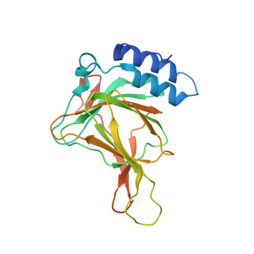
6N42

Crystal structure of cysteine-bound ferrous form of the crosslinked human cysteine dioxygenase in the anaerobic condition

  • Classification: OXIDOREDUCTASE
  • Organism(s): Homo sapiens
  • Expression System: Escherichia coli
  • Mutation(s): No 

  • Deposited: 2018-11-16 Released: 2019-04-17 
  • Deposition Author(s): Liu, A., Li, J.
  • Funding Organization(s): National Science Foundation (NSF, United States), National Institutes of Health/National Institute of General Medical Sciences (NIH/NIGMS)

Experimental Data Snapshot

  • Method: X-RAY DIFFRACTION
  • Resolution: 2.20 Å
  • R-Value Free: 0.210 
  • R-Value Work: 0.172 
  • R-Value Observed: 0.176 

Starting Model: experimental
View more details

wwPDB Validation   3D Report Full Report


Ligand Structure Quality Assessment 


This is version 1.5 of the entry. See complete history


Literature

Probing the Cys-Tyr Cofactor Biogenesis in Cysteine Dioxygenase by the Genetic Incorporation of Fluorotyrosine.

Li, J.Koto, T.Davis, I.Liu, A.

(2019) Biochemistry 58: 2218-2227

  • DOI: https://doi.org/10.1021/acs.biochem.9b00006
  • Primary Citation of Related Structures:  
    6BPR, 6E87, 6N42, 6N43

  • PubMed Abstract: 

    Cysteine dioxygenase (CDO) is a nonheme iron enzyme that adds two oxygen atoms from dioxygen to the sulfur atom of l-cysteine. Adjacent to the iron site of mammalian CDO, there is a post-translationally generated Cys-Tyr cofactor, whose presence substantially enhances the oxygenase activity. The formation of the Cys-Tyr cofactor in CDO is an autocatalytic process, and it is challenging to study by traditional techniques because the cross-linking reaction is a side, uncoupled, single-turnover oxidation buried among multiple turnovers of l-cysteine oxygenation. Here, we take advantage of our recent success in obtaining a purely uncross-linked human CDO due to site-specific incorporation of 3,5-difluoro-l-tyrosine (F 2 -Tyr) at the cross-linking site through the genetic code expansion strategy. Using EPR spectroscopy, we show that nitric oxide ( NO), an oxygen surrogate, similarly binds to uncross-linked F 2 -Tyr157 CDO as in wild-type human CDO. We determined X-ray crystal structures of uncross-linked F 2 -Tyr157 CDO and mature wild-type CDO in complex with both l-cysteine and NO. These structural data reveal that the active site cysteine (Cys93 in the human enzyme), rather than the generally expected tyrosine (i.e., Tyr157), is well-aligned to be oxidized should the normal oxidation reaction uncouple. This structure-based understanding is further supported by a computational study with models built on the uncross-linked ternary complex structure. Together, these results strongly suggest that the first target to oxidize during the iron-assisted Cys-Tyr cofactor biogenesis is Cys93. Based on these data, a plausible reaction mechanism implementing a cysteine radical involved in the cross-link formation is proposed.


  • Organizational Affiliation

    Department of Chemistry , University of Texas at San Antonio , San Antonio , Texas 78249 , United States.


Macromolecules
Find similar proteins by:  (by identity cutoff)  |  3D Structure
Entity ID: 1
MoleculeChains Sequence LengthOrganismDetailsImage
Cysteine dioxygenase type 1200Homo sapiensMutation(s): 0 
Gene Names: CDO1
EC: 1.13.11.20
UniProt & NIH Common Fund Data Resources
Find proteins for Q16878 (Homo sapiens)
Explore Q16878 
Go to UniProtKB:  Q16878
PHAROS:  Q16878
GTEx:  ENSG00000129596 
Entity Groups  
Sequence Clusters30% Identity50% Identity70% Identity90% Identity95% Identity100% Identity
UniProt GroupQ16878
Sequence Annotations
Expand
  • Reference Sequence
Small Molecules
Ligands 4 Unique
IDChains Name / Formula / InChI Key2D Diagram3D Interactions
CYS (Subject of Investigation/LOI)
Query on CYS

Download Ideal Coordinates CCD File 
C [auth A]CYSTEINE
C3 H7 N O2 S
XUJNEKJLAYXESH-REOHCLBHSA-N
SO4
Query on SO4

Download Ideal Coordinates CCD File 
I [auth A],
J [auth A],
K [auth A]
SULFATE ION
O4 S
QAOWNCQODCNURD-UHFFFAOYSA-L
GOL
Query on GOL

Download Ideal Coordinates CCD File 
D [auth A],
E [auth A],
F [auth A],
G [auth A],
H [auth A]
GLYCEROL
C3 H8 O3
PEDCQBHIVMGVHV-UHFFFAOYSA-N
FE2 (Subject of Investigation/LOI)
Query on FE2

Download Ideal Coordinates CCD File 
B [auth A]FE (II) ION
Fe
CWYNVVGOOAEACU-UHFFFAOYSA-N
Experimental Data & Validation

Experimental Data

  • Method: X-RAY DIFFRACTION
  • Resolution: 2.20 Å
  • R-Value Free: 0.210 
  • R-Value Work: 0.172 
  • R-Value Observed: 0.176 
  • Space Group: P 65
Unit Cell:
Length ( Å )Angle ( ˚ )
a = 131.221α = 90
b = 131.221β = 90
c = 34.296γ = 120
Software Package:
Software NamePurpose
DENZOdata reduction
SCALEPACKdata scaling
PHENIXrefinement
PDB_EXTRACTdata extraction
PHASERphasing

Structure Validation

View Full Validation Report


Ligand Structure Quality Assessment 


Entry History & Funding Information

Deposition Data

  • Released Date: 2019-04-17 
  • Deposition Author(s): Liu, A., Li, J.

Funding OrganizationLocationGrant Number
National Science Foundation (NSF, United States)United StatesCHE-1808637
National Institutes of Health/National Institute of General Medical Sciences (NIH/NIGMS)United StatesR01GM107529
National Institutes of Health/National Institute of General Medical Sciences (NIH/NIGMS)United StatesR01GM108988

Revision History  (Full details and data files)

  • Version 1.0: 2019-04-17
    Type: Initial release
  • Version 1.1: 2019-05-01
    Changes: Data collection, Database references
  • Version 1.2: 2019-05-15
    Changes: Data collection, Database references
  • Version 1.3: 2019-11-27
    Changes: Author supporting evidence
  • Version 1.4: 2023-10-11
    Changes: Data collection, Database references, Derived calculations, Refinement description
  • Version 1.5: 2024-10-23
    Changes: Structure summary