6N2G | pdb_00006n2g

Crystal structure of Caenorhabditis elegans NAP1


Experimental Data Snapshot

  • Method: X-RAY DIFFRACTION
  • Resolution: 3.00 Å
  • R-Value Free: 
    0.255 (Depositor), 0.260 (DCC) 
  • R-Value Work: 
    0.241 (Depositor), 0.247 (DCC) 
  • R-Value Observed: 
    0.242 (Depositor) 

wwPDB Validation 3D Report Full Report

Validation slider image for 6N2G

This is version 1.2 of the entry. See complete history

Literature

Characterization of Caenorhabditis elegans Nucleosome Assembly Protein 1 Uncovers the Role of Acidic Tails in Histone Binding.

Sarkar, P.Zhang, N.Bhattacharyya, S.Salvador, K.D'Arcy, S.

(2019) Biochemistry 58: 108-113

  • DOI: https://doi.org/10.1021/acs.biochem.8b01033
  • Primary Citation Related Structures: 
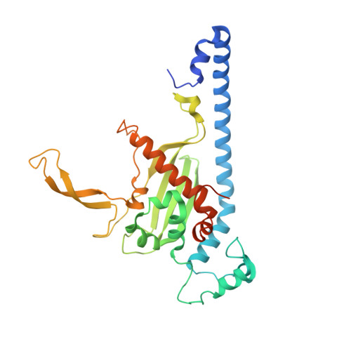
    6N2G

  • PubMed Abstract: 

    Nucleosome assembly proteins (Naps) influence chromatin dynamics by directly binding to histones. Here we provide a comprehensive structural and biochemical analysis of a Nap protein from Caenorhabditis elegans (CeNap1). CeNap1 naturally lacks the acidic N-terminal tail and has a short C-terminal tail compared to many other Nap proteins. Comparison of CeNap1 with full length and tail-less constructs of Saccharomyces cerevisiae Nap1 uncovers the role of these tails in self-association, histone binding, and Nap competition with DNA for H2A-H2B. We find that the presence of tails influences the stoichiometry of H2A-H2B binding and is required to complete the interactions between H2A-H2B and DNA. The absolute stoichiometry of the Nap protein and H2A-H2B complex is 2:1 or 2:2, with only a very small population of higher-order oligomers occurring at 150 mM NaCl. We also show that H3-H4 binds differently than H2A-H2B and that an (H3-H4) 2 tetramer can simultaneously bind two Nap 2 protein homodimers.


  • Organizational Affiliation
    • Department of Biochemistry and Molecular Biology , The University of Melbourne , Melbourne 3000 , Australia.

Macromolecule Content 

  • Total Structure Weight: 142.83 kDa 
  • Atom Count: 8,866 
  • Modeled Residue Count: 1,112 
  • Deposited Residue Count: 1,264 
  • Unique protein chains: 1

Macromolecules

Find similar proteins by:|  3D Structure
Entity ID: 1
MoleculeChains  Sequence LengthOrganismDetailsImage
Nucleosome Assembly Protein
A, B, C, D
316Caenorhabditis elegansMutation(s): 0 
Gene Names: nap-1CELE_D2096.8D2096.8
UniProt
Find proteins for Q19007 (Caenorhabditis elegans)
Explore Q19007 
Go to UniProtKB:  Q19007
Entity Groups
Sequence Clusters30% Identity50% Identity70% Identity90% Identity95% Identity100% Identity
UniProt GroupQ19007
Sequence Annotations
Expand
Reference Sequence

Experimental Data & Validation

Experimental Data

  • Method: X-RAY DIFFRACTION
  • Resolution: 3.00 Å
  • R-Value Free:  0.255 (Depositor), 0.260 (DCC) 
  • R-Value Work:  0.241 (Depositor), 0.247 (DCC) 
  • R-Value Observed: 0.242 (Depositor) 
Space Group: P 21 21 2
Unit Cell:
Length ( Å )Angle ( ˚ )
a = 125.597α = 90
b = 154.868β = 90
c = 92.551γ = 90
Software Package:
Software NamePurpose
PHENIXrefinement
SCALAdata scaling
PDB_EXTRACTdata extraction
XDSdata reduction
PHASERphasing

Structure Validation

View Full Validation Report


Entry History 

Deposition Data

Revision History  (Full details and data files)

  • Version 1.0: 2019-01-30
    Type: Initial release
  • Version 1.1: 2019-02-13
    Changes: Data collection, Database references
  • Version 1.2: 2024-03-13
    Changes: Data collection, Database references, Refinement description