6N1A | pdb_00006n1a

Crystal structure of an N-acetylgalactosamine deacetylase from F. plautii


Experimental Data Snapshot

  • Method: X-RAY DIFFRACTION
  • Resolution: 1.60 Å
  • R-Value Free: 
    0.167 (Depositor), 0.179 (DCC) 
  • R-Value Work: 
    0.139 (Depositor), 0.154 (DCC) 
  • R-Value Observed: 
    0.140 (Depositor) 

wwPDB Validation 3D Report Full Report

Validation slider image for 6N1A

This is version 1.4 of the entry. See complete history

Literature

An enzymatic pathway in the human gut microbiome that converts A to universal O type blood.

Rahfeld, P.Sim, L.Moon, H.Constantinescu, I.Morgan-Lang, C.Hallam, S.J.Kizhakkedathu, J.N.Withers, S.G.

(2019) Nat Microbiol 4: 1475-1485

  • DOI: https://doi.org/10.1038/s41564-019-0469-7
  • Primary Citation Related Structures: 
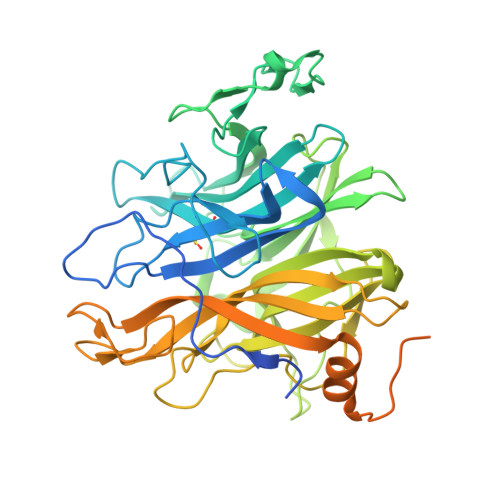
    6N1A, 6N1B

  • PubMed Abstract: 

    Access to efficient enzymes that can convert A and B type red blood cells to 'universal' donor O would greatly increase the supply of blood for transfusions. Here we report the functional metagenomic screening of the human gut microbiome for enzymes that can remove the cognate A and B type sugar antigens. Among the genes encoded in our library of 19,500 expressed fosmids bearing gut bacterial DNA, we identify an enzyme pair from the obligate anaerobe Flavonifractor plautii that work in concert to efficiently convert the A antigen to the H antigen of O type blood, via a galactosamine intermediate. The X-ray structure of the N-acetylgalactosamine deacetylase reveals the active site and mechanism of the founding member of an esterase family. The galactosaminidase expands activities within the CAZy family GH36. Their ability to completely convert A to O of the same rhesus type at very low enzyme concentrations in whole blood will simplify their incorporation into blood transfusion practice, broadening blood supply.


  • Organizational Affiliation
    • Department of Chemistry, University of British Columbia, Vancouver, British Columbia, Canada.

Macromolecule Content 

  • Total Structure Weight: 56.25 kDa 
  • Atom Count: 3,477 
  • Modeled Residue Count: 386 
  • Deposited Residue Count: 503 
  • Unique protein chains: 1

Macromolecules

Find similar proteins by:|  3D Structure
Entity ID: 1
MoleculeChains  Sequence LengthOrganismDetailsImage
Carbohydrate-binding protein503Flavonifractor plautiiMutation(s): 0 
Gene Names: A4U99_12080
UniProt
Find proteins for G9YSP4 (Flavonifractor plautii ATCC 29863)
Explore G9YSP4 
Go to UniProtKB:  G9YSP4
Entity Groups
Sequence Clusters30% Identity50% Identity70% Identity90% Identity95% Identity100% Identity
UniProt GroupG9YSP4
Sequence Annotations
Expand
Reference Sequence

Small Molecules

Modified Residues  1 Unique
IDChains TypeFormula2D DiagramParent
CSD
Query on CSD
A
L-PEPTIDE LINKINGC3 H7 N O4 SCYS

Experimental Data & Validation

Experimental Data

  • Method: X-RAY DIFFRACTION
  • Resolution: 1.60 Å
  • R-Value Free:  0.167 (Depositor), 0.179 (DCC) 
  • R-Value Work:  0.139 (Depositor), 0.154 (DCC) 
  • R-Value Observed: 0.140 (Depositor) 
Space Group: P 21 21 21
Unit Cell:
Length ( Å )Angle ( ˚ )
a = 51.584α = 90
b = 69.186β = 90
c = 104.325γ = 90
Software Package:
Software NamePurpose
REFMACrefinement
PDB_EXTRACTdata extraction
XDSdata reduction
Aimlessdata scaling
CRANK2phasing

Structure Validation

View Full Validation Report


Entry History 

& Funding Information

Deposition Data


Funding OrganizationLocationGrant Number
Canadian Institutes of Health Research (CIHR)CanadaMOP-136940

Revision History  (Full details and data files)

  • Version 1.0: 2019-06-12
    Type: Initial release
  • Version 1.1: 2019-07-10
    Changes: Data collection, Database references
  • Version 1.2: 2019-09-04
    Changes: Data collection, Database references
  • Version 1.3: 2020-01-08
    Changes: Author supporting evidence
  • Version 1.4: 2024-10-16
    Changes: Data collection, Database references, Derived calculations, Structure summary