6MYG | pdb_00006myg

Mouse Gamma S Crystallin L16 Octamer


Experimental Data Snapshot

  • Method: X-RAY DIFFRACTION
  • Resolution: 2.92 Å
  • R-Value Free: 
    0.284 (Depositor), 0.282 (DCC) 
  • R-Value Work: 
    0.205 (Depositor) 
  • R-Value Observed: 
    0.209 (Depositor) 

wwPDB Validation 3D Report Full Report

Validation slider image for 6MYG

This is version 1.1 of the entry. See complete history

Literature

Disulfides, domain swaps and disorder in a gamma-crystallin: A model for mechanisms of aggregation in globular proteins.

Sagar, V.Wistow, G.J.

To be published.

Macromolecule Content 

  • Total Structure Weight: 83.45 kDa 
  • Atom Count: 5,503 
  • Modeled Residue Count: 700 
  • Deposited Residue Count: 712 
  • Unique protein chains: 1

Macromolecules

Find similar proteins by:|  3D Structure
Entity ID: 1
MoleculeChains  Sequence LengthOrganismDetailsImage
Gamma-crystallin SA [auth B],
B [auth A],
C,
D
178Mus musculusMutation(s): 0 
Gene Names: Crygs
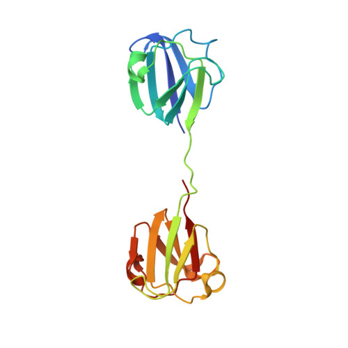
UniProt & NIH Common Fund Data Resources
Find proteins for O35486 (Mus musculus)
Explore O35486 
Go to UniProtKB:  O35486
IMPC:  MGI:1298216
Entity Groups
Sequence Clusters30% Identity50% Identity70% Identity90% Identity95% Identity100% Identity
UniProt GroupO35486
Sequence Annotations
Expand
Reference Sequence

Experimental Data & Validation

Experimental Data

  • Method: X-RAY DIFFRACTION
  • Resolution: 2.92 Å
  • R-Value Free:  0.284 (Depositor), 0.282 (DCC) 
  • R-Value Work:  0.205 (Depositor) 
  • R-Value Observed: 0.209 (Depositor) 
Space Group: P 21 21 2
Unit Cell:
Length ( Å )Angle ( ˚ )
a = 61.095α = 90
b = 77.591β = 90
c = 154.052γ = 90
Software Package:
Software NamePurpose
PHENIXrefinement
HKL-2000data reduction
HKL-2000data scaling
PHENIXphasing

Structure Validation

View Full Validation Report


Entry History 

Deposition Data

Revision History  (Full details and data files)

  • Version 1.0: 2018-11-14
    Type: Initial release
  • Version 1.1: 2024-11-13
    Changes: Data collection, Database references, Structure summary