6MYD | pdb_00006myd

Structure of zebrafish TRAF6 in complex with STING CTT


Experimental Data Snapshot

  • Method: X-RAY DIFFRACTION
  • Resolution: 1.40 Å
  • R-Value Free: 
    0.199 (Depositor), 0.200 (DCC) 
  • R-Value Work: 
    0.181 (Depositor), 0.184 (DCC) 
  • R-Value Observed: 
    0.181 (Depositor) 

Starting Model: experimental
View more details

wwPDB Validation 3D Report Full Report

Validation slider image for 6MYD

This is version 1.1 of the entry. See complete history

Literature

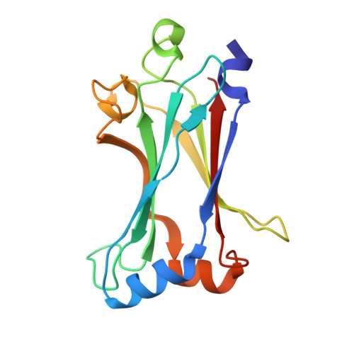
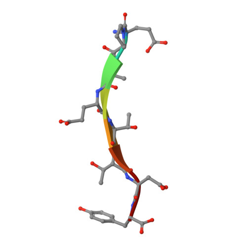
Modular Architecture of the STING C-Terminal Tail Allows Interferon and NF-kappa B Signaling Adaptation.

de Oliveira Mann, C.C.Orzalli, M.H.King, D.S.Kagan, J.C.Lee, A.S.Y.Kranzusch, P.J.

(2019) Cell Rep 27: 1165-1175.e5

  • DOI: https://doi.org/10.1016/j.celrep.2019.03.098
  • Primary Citation Related Structures: 
    6MYD

  • PubMed Abstract: 

    Stimulator of interferon genes (STING) is a key regulator of type I interferon and pro-inflammatory responses during infection, cellular stress, and cancer. Here, we reveal a mechanism for how STING balances activation of IRF3- and NF-κB-dependent transcription and discover that acquisition of discrete signaling modules in the vertebrate STING C-terminal tail (CTT) shapes downstream immunity. As a defining example, we identify a motif appended to the CTT of zebrafish STING that inverts the typical vertebrate signaling response and results in dramatic NF-κB activation and weak IRF3-interferon signaling. We determine a co-crystal structure that explains how this CTT sequence recruits TRAF6 as a new binding partner and demonstrate that the minimal motif is sufficient to reprogram human STING and immune activation in macrophage cells. Together, our results define the STING CTT as a linear signaling hub that can acquire modular motifs to readily adapt downstream immunity.


  • Organizational Affiliation
    • Department of Microbiology, Harvard Medical School, Boston, MA 02115, USA; Department of Cancer Immunology and Virology, Dana-Farber Cancer Institute, Boston, MA 02115, USA.

Macromolecule Content 

  • Total Structure Weight: 38.84 kDa 
  • Atom Count: 3,134 
  • Modeled Residue Count: 322 
  • Deposited Residue Count: 330 
  • Unique protein chains: 2

Macromolecules

Find similar proteins by:|  3D Structure
Entity ID: 1
MoleculeChains  Sequence LengthOrganismDetailsImage
TNF receptor-associated factor 6
A, C
157Danio rerioMutation(s): 0 
Gene Names: traf6si:dkey-56p7.3zgc:63704
EC: 2.3.2.27
UniProt
Find proteins for Q6IWL4 (Danio rerio)
Explore Q6IWL4 
Go to UniProtKB:  Q6IWL4
Entity Groups
Sequence Clusters30% Identity50% Identity70% Identity90% Identity95% Identity100% Identity
UniProt GroupQ6IWL4
Sequence Annotations
Expand
Reference Sequence
Find similar proteins by:  Sequence   |   3D Structure  
Entity ID: 2
MoleculeChains  Sequence LengthOrganismDetailsImage
STING CTT, Transmembrane protein 173
B, D
8Danio rerioMutation(s): 0 
UniProt
Find proteins for E7F4N7 (Danio rerio)
Explore E7F4N7 
Go to UniProtKB:  E7F4N7
Entity Groups
UniProt GroupE7F4N7
Sequence Annotations
Expand
Reference Sequence

Experimental Data & Validation

Experimental Data

  • Method: X-RAY DIFFRACTION
  • Resolution: 1.40 Å
  • R-Value Free:  0.199 (Depositor), 0.200 (DCC) 
  • R-Value Work:  0.181 (Depositor), 0.184 (DCC) 
  • R-Value Observed: 0.181 (Depositor) 
Space Group: P 1 21 1
Unit Cell:
Length ( Å )Angle ( ˚ )
a = 46.621α = 90
b = 84.377β = 115.42
c = 53.72γ = 90
Software Package:
Software NamePurpose
PHENIXrefinement
XDSdata reduction
Aimlessdata scaling
PHASERphasing

Structure Validation

View Full Validation Report


Entry History 

Deposition Data

Revision History  (Full details and data files)

  • Version 1.0: 2019-05-01
    Type: Initial release
  • Version 1.1: 2023-10-11
    Changes: Data collection, Database references, Refinement description