6MVL

Crystal structure of VISTA bound to a pH-selective antibody Fab fragment


Experimental Data Snapshot

  • Method: X-RAY DIFFRACTION
  • Resolution: 1.61 Å
  • R-Value Free: 
    0.225 (Depositor), 0.229 (DCC) 
  • R-Value Work: 
    0.182 (Depositor), 0.188 (DCC) 
  • R-Value Observed: 
    0.184 (Depositor) 

wwPDB Validation   3D Report Full Report


Ligand Structure Quality Assessment 

Created with Raphaël 2.3.0Worse 01 BetterLigand structure goodness of fit to experimental dataBest fitted NAGClick on this verticalbar to view details

This is version 1.3 of the entry. See complete history


Literature

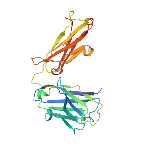
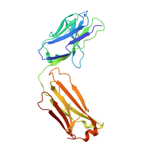
VISTA is an acidic pH-selective ligand for PSGL-1.

Johnston, R.J.Su, L.J.Pinckney, J.Critton, D.Boyer, E.Krishnakumar, A.Corbett, M.Rankin, A.L.Dibella, R.Campbell, L.Martin, G.H.Lemar, H.Cayton, T.Huang, R.Y.Deng, X.Nayeem, A.Chen, H.Ergel, B.Rizzo, J.M.Yamniuk, A.P.Dutta, S.Ngo, J.Shorts, A.O.Ramakrishnan, R.Kozhich, A.Holloway, J.Fang, H.Wang, Y.K.Yang, Z.Thiam, K.Rakestraw, G.Rajpal, A.Sheppard, P.Quigley, M.Bahjat, K.S.Korman, A.J.

(2019) Nature 574: 565-570

  • DOI: https://doi.org/10.1038/s41586-019-1674-5
  • Primary Citation of Related Structures:  
    6MVL

  • PubMed Abstract: 

    Co-inhibitory immune receptors can contribute to T cell dysfunction in patients with cancer 1,2 . Blocking antibodies against cytotoxic T-lymphocyte-associated protein 4 (CTLA-4) and programmed cell death 1 (PD-1) partially reverse this effect and are becoming standard of care in an increasing number of malignancies 3 . However, many of the other axes by which tumours become inhospitable to T cells are not fully understood. Here we report that V-domain immunoglobulin suppressor of T cell activation (VISTA) engages and suppresses T cells selectively at acidic pH such as that found in tumour microenvironments. Multiple histidine residues along the rim of the VISTA extracellular domain mediate binding to the adhesion and co-inhibitory receptor P-selectin glycoprotein ligand-1 (PSGL-1). Antibodies engineered to selectively bind and block this interaction in acidic environments were sufficient to reverse VISTA-mediated immune suppression in vivo. These findings identify a mechanism by which VISTA may engender resistance to anti-tumour immune responses, as well as an unexpectedly determinative role for pH in immune co-receptor engagement.


  • Organizational Affiliation

    Immuno-Oncology Discovery, Bristol-Myers Squibb, Redwood City, CA, USA. robert.johnston@bms.com.


Macromolecules
Find similar proteins by:  (by identity cutoff)  |  3D Structure
Entity ID: 1
MoleculeChains Sequence LengthOrganismDetailsImage
V-type immunoglobulin domain-containing suppressor of T-cell activation169Homo sapiensMutation(s): 3 
Gene Names: VSIRC10orf54SISP1VISTAPP2135UNQ730/PRO1412
UniProt & NIH Common Fund Data Resources
Find proteins for Q9H7M9 (Homo sapiens)
Explore Q9H7M9 
Go to UniProtKB:  Q9H7M9
PHAROS:  Q9H7M9
GTEx:  ENSG00000107738 
Entity Groups  
Sequence Clusters30% Identity50% Identity70% Identity90% Identity95% Identity100% Identity
UniProt GroupQ9H7M9
Glycosylation
Glycosylation Sites: 1Go to GlyGen: Q9H7M9-1
Sequence Annotations
Expand
  • Reference Sequence
Find similar proteins by:  (by identity cutoff)  |  3D Structure
Entity ID: 2
MoleculeChains Sequence LengthOrganismDetailsImage
Antibody Fab fragment heavy chainB [auth H]229Homo sapiensMutation(s): 0 
Entity Groups  
Sequence Clusters30% Identity50% Identity70% Identity90% Identity95% Identity100% Identity
Sequence Annotations
Expand
  • Reference Sequence
Find similar proteins by:  (by identity cutoff)  |  3D Structure
Entity ID: 3
MoleculeChains Sequence LengthOrganismDetailsImage
Antibody Fab fragment light chainC [auth L]215Homo sapiensMutation(s): 0 
Entity Groups  
Sequence Clusters30% Identity50% Identity70% Identity90% Identity95% Identity100% Identity
Sequence Annotations
Expand
  • Reference Sequence
Small Molecules
Ligands 2 Unique
IDChains Name / Formula / InChI Key2D Diagram3D Interactions
NAG
Query on NAG

Download Ideal Coordinates CCD File 
D [auth A]2-acetamido-2-deoxy-beta-D-glucopyranose
C8 H15 N O6
OVRNDRQMDRJTHS-FMDGEEDCSA-N
SO4
Query on SO4

Download Ideal Coordinates CCD File 
E [auth A]
F [auth A]
G [auth A]
H [auth A]
I [auth H]
SULFATE ION
O4 S
QAOWNCQODCNURD-UHFFFAOYSA-L
Experimental Data & Validation

Experimental Data

  • Method: X-RAY DIFFRACTION
  • Resolution: 1.61 Å
  • R-Value Free:  0.225 (Depositor), 0.229 (DCC) 
  • R-Value Work:  0.182 (Depositor), 0.188 (DCC) 
  • R-Value Observed: 0.184 (Depositor) 
Space Group: I 21 21 21
Unit Cell:
Length ( Å )Angle ( ˚ )
a = 66.166α = 90
b = 125.856β = 90
c = 192.04γ = 90
Software Package:
Software NamePurpose
BUSTERrefinement
XDSdata reduction
XDSdata scaling
PHASERphasing

Structure Validation

View Full Validation Report


Ligand Structure Quality Assessment 

Created with Raphaël 2.3.0Worse 01 BetterLigand structure goodness of fit to experimental dataBest fitted NAGClick on this verticalbar to view details

Entry History 

Deposition Data

Revision History  (Full details and data files)

  • Version 1.0: 2019-10-23
    Type: Initial release
  • Version 1.1: 2019-11-06
    Changes: Data collection, Database references
  • Version 1.2: 2020-07-29
    Type: Remediation
    Reason: Carbohydrate remediation
    Changes: Data collection, Derived calculations, Structure summary
  • Version 1.3: 2024-10-23
    Changes: Data collection, Database references, Structure summary