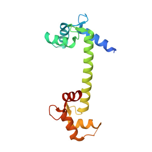
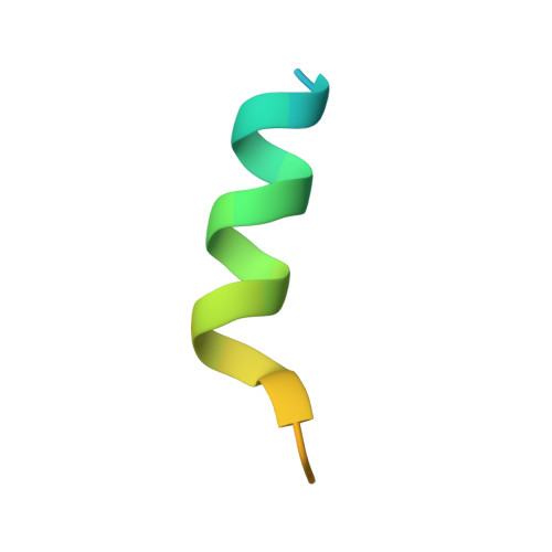
6MUE

Voltage-gated sodium channel NaV1.4 IQ domain in complex with Ca2+/Calmodulin


Experimental Data Snapshot

  • Method: X-RAY DIFFRACTION
  • Resolution: 1.90 Å
  • R-Value Free: 0.260 
  • R-Value Work: 0.214 
  • R-Value Observed: 0.216 

Starting Model: experimental
View more details

wwPDB Validation   3D Report Full Report


This is version 1.5 of the entry. See complete history


Literature

Crystal structures of Ca2+-calmodulin bound to NaVC-terminal regions suggest role for EF-hand domain in binding and inactivation.

Gardill, B.R.Rivera-Acevedo, R.E.Tung, C.C.Van Petegem, F.

(2019) Proc Natl Acad Sci U S A 116: 10763-10772

  • DOI: https://doi.org/10.1073/pnas.1818618116
  • Primary Citation of Related Structures:  
    6MUD, 6MUE

  • PubMed Abstract: 

    Voltage-gated sodium (Na V ) and calcium channels (Ca V ) form targets for calmodulin (CaM), which affects channel inactivation properties. A major interaction site for CaM resides in the C-terminal (CT) region, consisting of an IQ domain downstream of an EF-hand domain. We present a crystal structure of fully Ca 2+ -occupied CaM, bound to the CT of Na V 1.5. The structure shows that the C-terminal lobe binds to a site ∼90° rotated relative to a previous site reported for an apoCaM complex with the Na V 1.5 CT and for ternary complexes containing fibroblast growth factor homologous factors (FHF). We show that the binding of FHFs forces the EF-hand domain in a conformation that does not allow binding of the Ca 2+ -occupied C-lobe of CaM. These observations highlight the central role of the EF-hand domain in modulating the binding mode of CaM. The binding sites for Ca 2+ -free and Ca 2+ -occupied CaM contain targets for mutations linked to long-QT syndrome, a type of inherited arrhythmia. The related Na V 1.4 channel has been shown to undergo Ca 2+ -dependent inactivation (CDI) akin to Ca V s. We present a crystal structure of Ca 2+ /CaM bound to the Na V 1.4 IQ domain, which shows a binding mode that would clash with the EF-hand domain. We postulate the relative reorientation of the EF-hand domain and the IQ domain as a possible conformational switch that underlies CDI.


  • Organizational Affiliation

    Department of Biochemistry and Molecular Biology, Life Sciences Institute, The University of British Columbia, Vancouver, BC, Canada V6T 1Z3.


Macromolecules
Find similar proteins by:  (by identity cutoff)  |  3D Structure
Entity ID: 1
MoleculeChains Sequence LengthOrganismDetailsImage
Calmodulin-1151Homo sapiensMutation(s): 0 
Gene Names: CALM1CALMCAMCAM1
UniProt & NIH Common Fund Data Resources
Find proteins for P0DP23 (Homo sapiens)
Explore P0DP23 
Go to UniProtKB:  P0DP23
PHAROS:  P0DP23
GTEx:  ENSG00000198668 
Entity Groups  
Sequence Clusters30% Identity50% Identity70% Identity90% Identity95% Identity100% Identity
UniProt GroupP0DP23
Sequence Annotations
Expand
  • Reference Sequence
Find similar proteins by:  (by identity cutoff)  |  3D Structure
Entity ID: 2
MoleculeChains Sequence LengthOrganismDetailsImage
Sodium channel protein type 4 subunit alpha29Rattus norvegicusMutation(s): 0 
UniProt
Find proteins for P15390 (Rattus norvegicus)
Explore P15390 
Go to UniProtKB:  P15390
Entity Groups  
Sequence Clusters30% Identity50% Identity70% Identity90% Identity95% Identity100% Identity
UniProt GroupP15390
Sequence Annotations
Expand
  • Reference Sequence
Experimental Data & Validation

Experimental Data

  • Method: X-RAY DIFFRACTION
  • Resolution: 1.90 Å
  • R-Value Free: 0.260 
  • R-Value Work: 0.214 
  • R-Value Observed: 0.216 
  • Space Group: P 1 2 1
Unit Cell:
Length ( Å )Angle ( ˚ )
a = 58.58α = 90
b = 24.433β = 112.63
c = 60.72γ = 90
Software Package:
Software NamePurpose
REFMACrefinement
PDB_EXTRACTdata extraction
XDSdata reduction
XDSdata scaling
PHASERphasing

Structure Validation

View Full Validation Report


Entry History & Funding Information

Deposition Data


Funding OrganizationLocationGrant Number
Canadian Institutes of Health Research (CIHR)CanadaMOP-119404

Revision History  (Full details and data files)

  • Version 1.0: 2019-05-08
    Type: Initial release
  • Version 1.1: 2019-05-22
    Changes: Data collection, Database references
  • Version 1.2: 2019-06-12
    Changes: Data collection, Database references
  • Version 1.3: 2020-01-08
    Changes: Author supporting evidence
  • Version 1.4: 2023-10-11
    Changes: Data collection, Database references, Refinement description
  • Version 1.5: 2024-11-20
    Changes: Structure summary