6MMO | pdb_00006mmo

Carbon regulatory PII-like protein SbtB from Cyanobium sp. 7001 bound to AMP


Experimental Data Snapshot

  • Method: X-RAY DIFFRACTION
  • Resolution: 1.86 Å
  • R-Value Free: 
    0.252 (Depositor), 0.255 (DCC) 
  • R-Value Work: 
    0.210 (Depositor), 0.212 (DCC) 
  • R-Value Observed: 
    0.212 (Depositor) 

wwPDB Validation 3D Report Full Report

Validation slider image for 6MMO

Ligand Structure Quality Assessment 


This is version 1.1 of the entry. See complete history

Literature

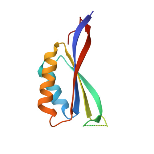
Structure and function of SbtB from Cyanobium sp. 7001

Jackson, C.Kaczmarski, J.A.Price, D.

(2019) bioRxiv 

Macromolecule Content 

  • Total Structure Weight: 71.19 kDa 
  • Atom Count: 4,337 
  • Modeled Residue Count: 524 
  • Deposited Residue Count: 624 
  • Unique protein chains: 1

Macromolecules

Find similar proteins by:|  3D Structure
Entity ID: 1
MoleculeChains  Sequence LengthOrganismDetailsImage
Carbon regulatory PII-like protein SbtB
A, B, C, D, E
A, B, C, D, E, F
104Cyanobium sp. PCC 7001Mutation(s): 0 
Gene Names: CPCC7001_1671
UniProt
Find proteins for A0ACD6B9N9 (Cyanobium sp. PCC 7001)
Explore A0ACD6B9N9 
Go to UniProtKB:  A0ACD6B9N9
Entity Groups
Sequence Clusters30% Identity50% Identity70% Identity90% Identity95% Identity100% Identity
UniProt GroupA0ACD6B9N9
Sequence Annotations
Expand
Reference Sequence

Small Molecules

Ligands 3 Unique
IDChains Name / Formula / InChI Key2D Diagram3D Interactions
AMP
(Subject of Investigation/LOI)

Query on AMP


Download:Ideal Coordinates CCD File
K [auth B]
N [auth C]
O [auth D]
P [auth D]
Q [auth E]
K [auth B],
N [auth C],
O [auth D],
P [auth D],
Q [auth E],
S [auth F]
ADENOSINE MONOPHOSPHATE
C10 H14 N5 O7 P
UDMBCSSLTHHNCD-KQYNXXCUSA-N
CL

Query on CL


Download:Ideal Coordinates CCD File
G [auth A]CHLORIDE ION
Cl
VEXZGXHMUGYJMC-UHFFFAOYSA-M
MG

Query on MG


Download:Ideal Coordinates CCD File
H [auth A]
I [auth A]
J [auth A]
L [auth B]
M [auth B]
H [auth A],
I [auth A],
J [auth A],
L [auth B],
M [auth B],
R [auth E],
T [auth F]
MAGNESIUM ION
Mg
JLVVSXFLKOJNIY-UHFFFAOYSA-N

Experimental Data & Validation

Experimental Data

  • Method: X-RAY DIFFRACTION
  • Resolution: 1.86 Å
  • R-Value Free:  0.252 (Depositor), 0.255 (DCC) 
  • R-Value Work:  0.210 (Depositor), 0.212 (DCC) 
  • R-Value Observed: 0.212 (Depositor) 
Space Group: P 1
Unit Cell:
Length ( Å )Angle ( ˚ )
a = 46.737α = 97.91
b = 51.332β = 102.55
c = 66.803γ = 103.09
Software Package:
Software NamePurpose
PHENIXrefinement
XDSdata reduction
Aimlessdata scaling
PHASERphasing

Structure Validation

View Full Validation Report


Ligand Structure Quality Assessment 


Entry History 

Deposition Data

Revision History  (Full details and data files)

  • Version 1.0: 2019-09-18
    Type: Initial release
  • Version 1.1: 2024-03-13
    Changes: Data collection, Database references, Derived calculations