


X-ray crystal structure of darunavir-resistant-P51 HIV-1 protease in complex with GRL-121
Yedidi, R.S., Hayashi, H., Das, D., Mitsuya, H.To be published.
Experimental Data Snapshot
Starting Model: experimental
View more details
Entity ID: 1 | |||||
|---|---|---|---|---|---|
| Molecule | Chains | Sequence Length | Organism | Details | Image |
| Protease | 99 | Human immunodeficiency virus 1 | Mutation(s): 0 Gene Names: pol | 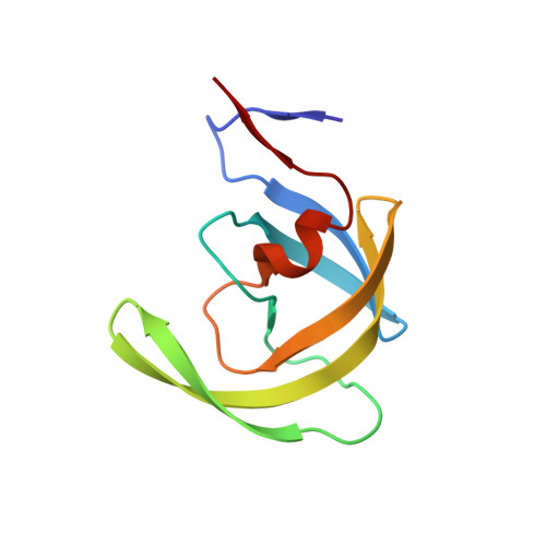 | |
UniProt | |||||
Find proteins for A0A4P8EW36 (Human immunodeficiency virus type 1) Explore A0A4P8EW36 Go to UniProtKB: A0A4P8EW36 | |||||
Entity Groups | |||||
| Sequence Clusters | 30% Identity50% Identity70% Identity90% Identity95% Identity100% Identity | ||||
| UniProt Group | A0A4P8EW36 | ||||
Sequence AnnotationsExpand | |||||
Reference Sequence | |||||
| Ligands 1 Unique | |||||
|---|---|---|---|---|---|
| ID | Chains | Name / Formula / InChI Key | 2D Diagram | 3D Interactions | |
| 7O7 Download:Ideal Coordinates CCD File | C [auth A] | (3S,3aR,5R,7aS,8S)-hexahydro-4H-3,5-methanofuro[2,3-b]pyran-8-yl {(2S,3R)-4-[{[2-(cyclopropylamino)-1,3-benzothiazol-6-yl]sulfonyl}(2-methylpropyl)amino]-3-hydroxy-1-phenylbutan-2-yl}carbamate C33 H42 N4 O7 S2 VXTSDGOABSRKIR-BLFKHSGCSA-N |  | ||
| Length ( Å ) | Angle ( ˚ ) |
|---|---|
| a = 62.799 | α = 90 |
| b = 62.799 | β = 90 |
| c = 82.806 | γ = 120 |
| Software Name | Purpose |
|---|---|
| PHENIX | refinement |
| HKL-2000 | data reduction |
| HKL-2000 | data scaling |
| MOLREP | phasing |
| Funding Organization | Location | Grant Number |
|---|---|---|
| National Institutes of Health/National Cancer Institute (NIH/NCI) | United States | -- |


