6MI6 | pdb_00006mi6

STRUCTURE OF CHEA DOMAIN P4 IN COMPLEX WITH AN ADP ANALOG


Experimental Data Snapshot

  • Method: X-RAY DIFFRACTION
  • Resolution: 2.95 Å
  • R-Value Free: 
    0.298 (Depositor), 0.292 (DCC) 
  • R-Value Work: 
    0.233 (Depositor), 0.229 (DCC) 
  • R-Value Observed: 
    0.240 (Depositor) 

wwPDB Validation 3D Report Full Report

Validation slider image for 6MI6

Ligand Structure Quality Assessment 


This is version 1.2 of the entry. See complete history

Literature

Nucleotide Spin Labeling for ESR Spectroscopy of ATP-Binding Proteins.

Muok, A.R.Chua, T.K.Le, H.Crane, B.R.

(2018) Appl Magn Reson 49: 1385-1395

  • DOI: https://doi.org/10.1007/s00723-018-1070-6
  • Primary Citation Related Structures: 
    6MI6

  • PubMed Abstract: 

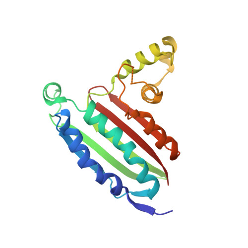
    Site-directed spin labeling of proteins by chemical modification of engineered cysteine residues with the molecule MTSSL (1-Oxyl-2,2,5,5-tetramethylpyrroline-3-methyl methanethiosulfonate) has been an invaluable tool for conducting double electron electron resonance (DEER) spectroscopy experiments. However, this method is generally limited to recombinant proteins with a limited number of reactive Cys residues that when modified will not impair protein function. Here we present a method that allows for spin-labeling of protein nucleotide binding sites by adenosine diphosphate (ADP) modified with a nitroxide moiety on the β-phosphate (ADP-β-S-SL). The synthesis of this ADP analog is straightforward and isolation of pure product is readily achieved on a standard reverse-phase high-performance liquid chromatography (HPLC) system. Furthermore, analyses of isolated ADP-β-S-SL by LC-mass spectrometry confirm that the molecule is extremely stable under ambient conditions. The crystal structure of ADP-β-S-SL bound to the ATP pocket of the histidine kinase CheA reveals specific targeting of the probe, whose nitroxide moiety is mobile on the protein surface. Continuous wave and pulsed ESR measurements demonstrate the capability of ADP-β-S-SL to report on active site environment and provide reliable DEER distance constraints.


  • Organizational Affiliation
    • Department of Chemistry and Chemical Biology, Cornell University Ithaca, NY.

Macromolecule Content 

  • Total Structure Weight: 86.36 kDa 
  • Atom Count: 5,687 
  • Modeled Residue Count: 729 
  • Deposited Residue Count: 748 
  • Unique protein chains: 1

Macromolecules

Find similar proteins by:|  3D Structure
Entity ID: 1
MoleculeChains  Sequence LengthOrganismDetailsImage
Chemotaxis protein CheA
A, B, C, D
187Thermotoga maritima MSB8Mutation(s): 0 
Gene Names: cheATM_0702
EC: 2.7.13.3
UniProt
Find proteins for Q56310 (Thermotoga maritima (strain ATCC 43589 / DSM 3109 / JCM 10099 / NBRC 100826 / MSB8))
Explore Q56310 
Go to UniProtKB:  Q56310
Entity Groups
Sequence Clusters30% Identity50% Identity70% Identity90% Identity95% Identity100% Identity
UniProt GroupQ56310
Sequence Annotations
Expand
Reference Sequence

Small Molecules

Ligands 3 Unique
IDChains Name / Formula / InChI Key2D Diagram3D Interactions
JSJ

Query on JSJ


Download:Ideal Coordinates CCD File
F [auth A],
H [auth B]
5'-O-[(S)-hydroxy{[(S)-hydroxy{[(1-hydroxy-2,2,5,5-tetramethyl-2,5-dihydro-1H-pyrrol-3-yl)methyl]disulfanyl}phosphoryl]oxy}phosphoryl]adenosine
C19 H30 N6 O10 P2 S2
FXTBVPRAZDKBSQ-LSCFUAHRSA-N
ADP

Query on ADP


Download:Ideal Coordinates CCD File
J [auth C],
L [auth D]
ADENOSINE-5'-DIPHOSPHATE
C10 H15 N5 O10 P2
XTWYTFMLZFPYCI-KQYNXXCUSA-N
SO4

Query on SO4


Download:Ideal Coordinates CCD File
E [auth A],
G [auth A],
I [auth C],
K [auth C]
SULFATE ION
O4 S
QAOWNCQODCNURD-UHFFFAOYSA-L

Experimental Data & Validation

Experimental Data

  • Method: X-RAY DIFFRACTION
  • Resolution: 2.95 Å
  • R-Value Free:  0.298 (Depositor), 0.292 (DCC) 
  • R-Value Work:  0.233 (Depositor), 0.229 (DCC) 
  • R-Value Observed: 0.240 (Depositor) 
Space Group: P 1 21 1
Unit Cell:
Length ( Å )Angle ( ˚ )
a = 83.703α = 90
b = 75.145β = 90.01
c = 86.58γ = 90
Software Package:
Software NamePurpose
PHENIXrefinement
HKL-2000data collection
PHASERphasing
Cootmodel building

Structure Validation

View Full Validation Report


Ligand Structure Quality Assessment 


Entry History 

Deposition Data

Revision History  (Full details and data files)

  • Version 1.0: 2018-12-05
    Type: Initial release
  • Version 1.1: 2019-11-13
    Changes: Database references
  • Version 1.2: 2024-12-25
    Changes: Advisory, Data collection, Database references, Derived calculations, Structure summary