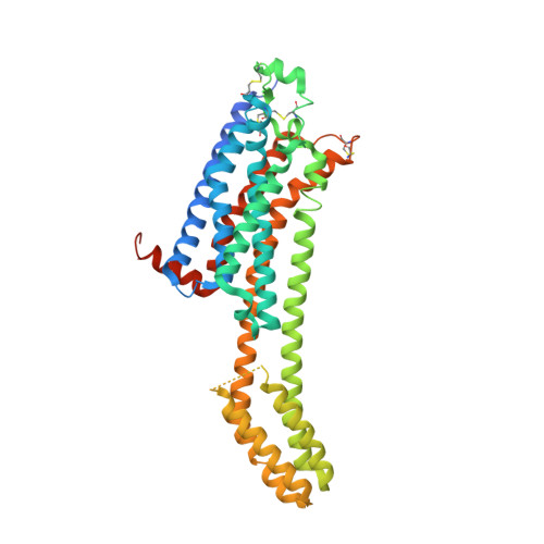
6MH8

High-viscosity injector-based Pink Beam Serial Crystallography of Micro-crystals at a Synchrotron Radiation Source


Experimental Data Snapshot

  • Method: X-RAY DIFFRACTION
  • Resolution: 4.20 Å
  • R-Value Free: 
    0.288 (Depositor), 0.280 (DCC) 
  • R-Value Work: 
    0.250 (Depositor), 0.250 (DCC) 
  • R-Value Observed: 
    0.254 (Depositor) 

Starting Model: experimental
View more details

wwPDB Validation   3D Report Full Report


Ligand Structure Quality Assessment 

Created with Raphaël 2.3.0Worse 01 BetterLigand structure goodness of fit to experimental dataBest fitted ZMAClick on this verticalbar to view details

This is version 1.7 of the entry. See complete history


Literature

High-viscosity injector-based pink-beam serial crystallography of microcrystals at a synchrotron radiation source.

Martin-Garcia, J.M.Zhu, L.Mendez, D.Lee, M.Y.Chun, E.Li, C.Hu, H.Subramanian, G.Kissick, D.Ogata, C.Henning, R.Ishchenko, A.Dobson, Z.Zhang, S.Weierstall, U.Spence, J.C.H.Fromme, P.Zatsepin, N.A.Fischetti, R.F.Cherezov, V.Liu, W.

(2019) IUCrJ 6: 412-425

  • DOI: https://doi.org/10.1107/S205225251900263X
  • Primary Citation of Related Structures:  
    6MH6, 6MH8

  • PubMed Abstract: 

    Since the first successful serial crystallography (SX) experiment at a synchrotron radiation source, the popularity of this approach has continued to grow showing that third-generation synchrotrons can be viable alternatives to scarce X-ray free-electron laser sources. Synchrotron radiation flux may be increased ∼100 times by a moderate increase in the bandwidth ('pink beam' conditions) at some cost to data analysis complexity. Here, we report the first high-viscosity injector-based pink-beam SX experiments. The structures of proteinase K (PK) and A 2A adenosine receptor (A 2A AR) were determined to resolutions of 1.8 and 4.2 Å using 4 and 24 consecutive 100 ps X-ray pulse exposures, respectively. Strong PK data were processed using existing Laue approaches, while weaker A 2A AR data required an alternative data-processing strategy. This demonstration of the feasibility presents new opportunities for time-resolved experiments with microcrystals to study structural changes in real time at pink-beam synchrotron beamlines worldwide.


  • Organizational Affiliation

    Biodesign Center for Applied Structural Discovery, Biodesign Institute, Arizona State University, 727 East Tyler Street, Tempe, AZ 85287, USA.


Macromolecules
Find similar proteins by:  (by identity cutoff)  |  3D Structure
Entity ID: 1
MoleculeChains Sequence LengthOrganismDetailsImage
Adenosine receptor A2a, Soluble cytochrome b562 chimeric construct447Homo sapiensEscherichia coliMutation(s): 0 
Gene Names: ADORA2AADORA2cybC
Membrane Entity: Yes 
UniProt & NIH Common Fund Data Resources
Find proteins for P0ABE7 (Escherichia coli)
Explore P0ABE7 
Go to UniProtKB:  P0ABE7
Find proteins for P29274 (Homo sapiens)
Explore P29274 
Go to UniProtKB:  P29274
PHAROS:  P29274
GTEx:  ENSG00000128271 
Entity Groups  
Sequence Clusters30% Identity50% Identity70% Identity90% Identity95% Identity100% Identity
UniProt GroupsP0ABE7P29274
Sequence Annotations
Expand
  • Reference Sequence
Small Molecules
Ligands 1 Unique
IDChains Name / Formula / InChI Key2D Diagram3D Interactions
ZMA
Query on ZMA

Download Ideal Coordinates CCD File 
B [auth A]4-{2-[(7-amino-2-furan-2-yl[1,2,4]triazolo[1,5-a][1,3,5]triazin-5-yl)amino]ethyl}phenol
C16 H15 N7 O2
PWTBZOIUWZOPFT-UHFFFAOYSA-N
Binding Affinity Annotations 
IDSourceBinding Affinity
ZMA BindingDB:  6MH8 Ki: min: 0.1, max: 395 (nM) from 18 assay(s)
Kd: min: 0.22, max: 286 (nM) from 2 assay(s)
IC50: min: 0.68, max: 81 (nM) from 7 assay(s)
Experimental Data & Validation

Experimental Data

  • Method: X-RAY DIFFRACTION
  • Resolution: 4.20 Å
  • R-Value Free:  0.288 (Depositor), 0.280 (DCC) 
  • R-Value Work:  0.250 (Depositor), 0.250 (DCC) 
  • R-Value Observed: 0.254 (Depositor) 
Space Group: C 2 2 21
Unit Cell:
Length ( Å )Angle ( ˚ )
a = 40α = 90
b = 179β = 90
c = 142γ = 90
Software Package:
Software NamePurpose
REFMACrefinement
CrystFELdata reduction
PHASERphasing

Structure Validation

View Full Validation Report


Ligand Structure Quality Assessment 

Created with Raphaël 2.3.0Worse 01 BetterLigand structure goodness of fit to experimental dataBest fitted ZMAClick on this verticalbar to view details

Entry History & Funding Information

Deposition Data


Funding OrganizationLocationGrant Number
National Science Foundation (NSF, United States)United States1231306
National Science Foundation (NSF, United States)United States1565180
National Institutes of Health/National Institute of General Medical Sciences (NIH/NIGMS)United StatesR21DA042298
National Institutes of Health/National Institute of General Medical Sciences (NIH/NIGMS)United StatesR01GM124152
National Institutes of Health/National Institute of General Medical Sciences (NIH/NIGMS)United StatesR01GM095583
National Institutes of Health/National Institute of General Medical Sciences (NIH/NIGMS)United StatesR35GM127086
National Institutes of Health/National Institute of General Medical Sciences (NIH/NIGMS)United StatesR24GM111072
National Institutes of Health/National Cancer Institute (NIH/NCI)United StatesACB-12002
National Institutes of Health/National Institute of General Medical Sciences (NIH/NIGMS)United StatesAGM12006
Department of Energy (DOE, United States)United StatesDE-AC02-06CH11357

Revision History  (Full details and data files)

  • Version 1.0: 2019-04-24
    Type: Initial release
  • Version 1.1: 2019-05-29
    Changes: Data collection, Database references
  • Version 1.2: 2019-08-21
    Changes: Data collection
  • Version 1.3: 2019-11-06
    Changes: Author supporting evidence, Data collection
  • Version 1.4: 2019-11-27
    Changes: Author supporting evidence
  • Version 1.5: 2023-10-11
    Changes: Data collection, Database references, Refinement description
  • Version 1.6: 2023-11-01
    Changes: Refinement description
  • Version 1.7: 2024-11-20
    Changes: Structure summary