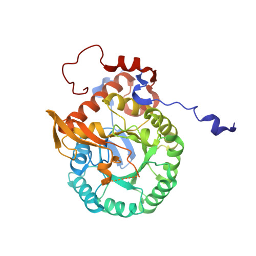
Crystal Structure of the Catalytic Domain of the Inosine Monophosphate Dehydrogenase from Bacillus Anthracis in the complex with inhibitor Oxanosine monophosphate
Kim, Y., Maltseva, N., Yu, R., Hedstrom, L., Joachimiak, A., Center for Structural Genomics of Infectious Diseases (CSGID)To be published.